大家都在搜

手机验证

询价列表

暂时没有已询价产品
Recombinant protein encompassing a sequence within the center region of human ER81. The exact sequence is proprietary.
IgG
Liquid
Store as concentrated solution. Centrifuge briefly prior to opening vial. For short-term storage (1-2 weeks), store at 4ºC. For long-term storage, aliquot and store at -20ºC or below. Avoid multiple freeze-thaw cycles.
Polyclonal
Unconjugated
Human, Mouse, Rat
12 months from the shipping date of the product.
Human
GTX129202
Primary Antibodies
Available
GeneTex
Rabbit
WB, ICC/IF, IHC-P, IHC-Fr
0.83 mg/ml (Please refer to the vial label for the specific concentration.)
This antibody was rasied against ER81 isoform 1. Based on sequence homology, this antibody can recognize most ER81 isoforms, but it may be unable to recognize isoform 4 according to the customer's feedback.
ER81 antibody
ER81 抗体
100 μl/25 μl
| 规格: | 100 μl | 产品价格: | ¥4000.0 |
|---|---|---|---|
| 规格: | 25 μl | 产品价格: | ¥1700.0 |

ER81 antibody detects ER81 protein at nucleus by immunofluorescent analysis.Sample: U87-MG cells were fixed in 4% PFA at RT for 15 min.Green: ER81 stained by ER81 antibody (GTX129202) diluted at 1:200.Red: alpha Tubulin, a cytoskeleton marker, stained by alpha Tubulin antibody [GT114] (GTX628802) diluted at 1:1000.Blue: Fluoroshield with DAPI (GTX30920).Scale bar= 10μm.

ER81 antibody detects ER81 protein by immunofluorescent analysis.Sample: DIV9 rat hippocampal neuron and Glia cell cells were fixed in 4% PFA at RT for 15 min.Green: ER81 stained by ER81 antibody (GTX129202) diluted at 1:250.Red: Tau, a Axon marker, stained by Tau antibody [GT287] (GTX634809) diluted at 1:500.

ER81 antibody detects ER81 protein at nucleus by immunohistochemical analysis.Sample: Paraffin-embedded rat brain.ER81 stained by ER81 antibody (GTX129202) diluted at 1:500.Antigen Retrieval: Citrate buffer, pH 6.0, 15 min

ER81 antibody detects ER81 protein at nucleus by immunohistochemical analysis.Sample: Paraffin-embedded rat brain.ER81 stained by ER81 antibody (GTX129202) diluted at 1:500.Antigen Retrieval: Citrate buffer, pH 6.0, 15 min

Non-transfected (–) and transfected (+) 293T whole cell extracts (30 μg) were separated by 10% SDS-PAGE, and the membrane was blotted with ER81 antibody (GTX129202) diluted at 1:5000. The HRP-conjugated anti-rabbit IgG antibody (GTX213110-01) was used to detect the primary antibody.

ER81 antibody detects ER81 protein at nucleus by immunohistochemical analysis.Sample: Paraffin-embedded mouse brain.ER81 stained by ER81 antibody (GTX129202) diluted at 1:500.Antigen Retrieval: Citrate buffer, pH 6.0, 15 min

ER81 antibody detects ER81 protein by immunohistochemical analysis.Sample: Frozen-sectioned mouse cerebellum.Green: ER81 stained by ER81 antibody (GTX129202) diluted at 1:250.Red: NF-H, stained by NF-H antibody [GT114] (GTX634289) diluted at 1:500.Blue: Fluoroshield with DAPI (GTX30920).

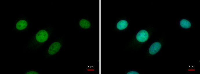
ER81 antibody detects ER81 protein at nucleus by immunofluorescent analysis.
Sample: C8D30 cells were fixed in 4% PFA at RT for 15 min.
Green: ER81 protein stained by ER81 antibody (GTX129202) diluted at 1:500.
Blue: Hoechst 33342 staining.

ER81 antibody detects ER81 protein at nucleus by immunohistochemical analysis.Sample: Paraffin-embedded mouse brain.ER81 stained by ER81 antibody (GTX129202) diluted at 1:500.Antigen Retrieval: Citrate buffer, pH 6.0, 15 min

U87-MG whole cell and nuclear extracts (30 μg) were separated by 10% SDS-PAGE, and the membrane was blotted with ER81 antibody (GTX129202) diluted at 1:1000. The HRP-conjugated anti-rabbit IgG antibody (GTX213110-01) was used to detect the primary antibody.

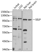
Whole cell extract (30 μg) was separated by 10% SDS-PAGE, and the membrane was blotted with ER81 antibody (GTX129202) diluted at 1:1000. The HRP-conjugated anti-rabbit IgG antibody (GTX213110-01) was used to detect the primary antibody.
风险提示:丁香通仅作为第三方平台,为商家信息发布提供平台空间。用户咨询产品时请注意保护个人信息及财产安全,合理判断,谨慎选购商品,商家和用户对交易行为负责。对于医疗器械类产品,请先查证核实企业经营资质和医疗器械产品注册证情况。