

- 询价
- YSKD
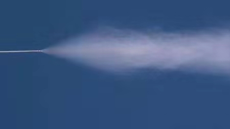
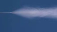
- 大鼠呼吸道定量给药器,大鼠气管内定量给药器
- 2026年04月30日

企业认证
相关产品推荐更多 >


万千商家帮你免费找货
0 人在求购买到急需产品
- 详细信息
- 询价记录
- 文献和实验
- 技术资料
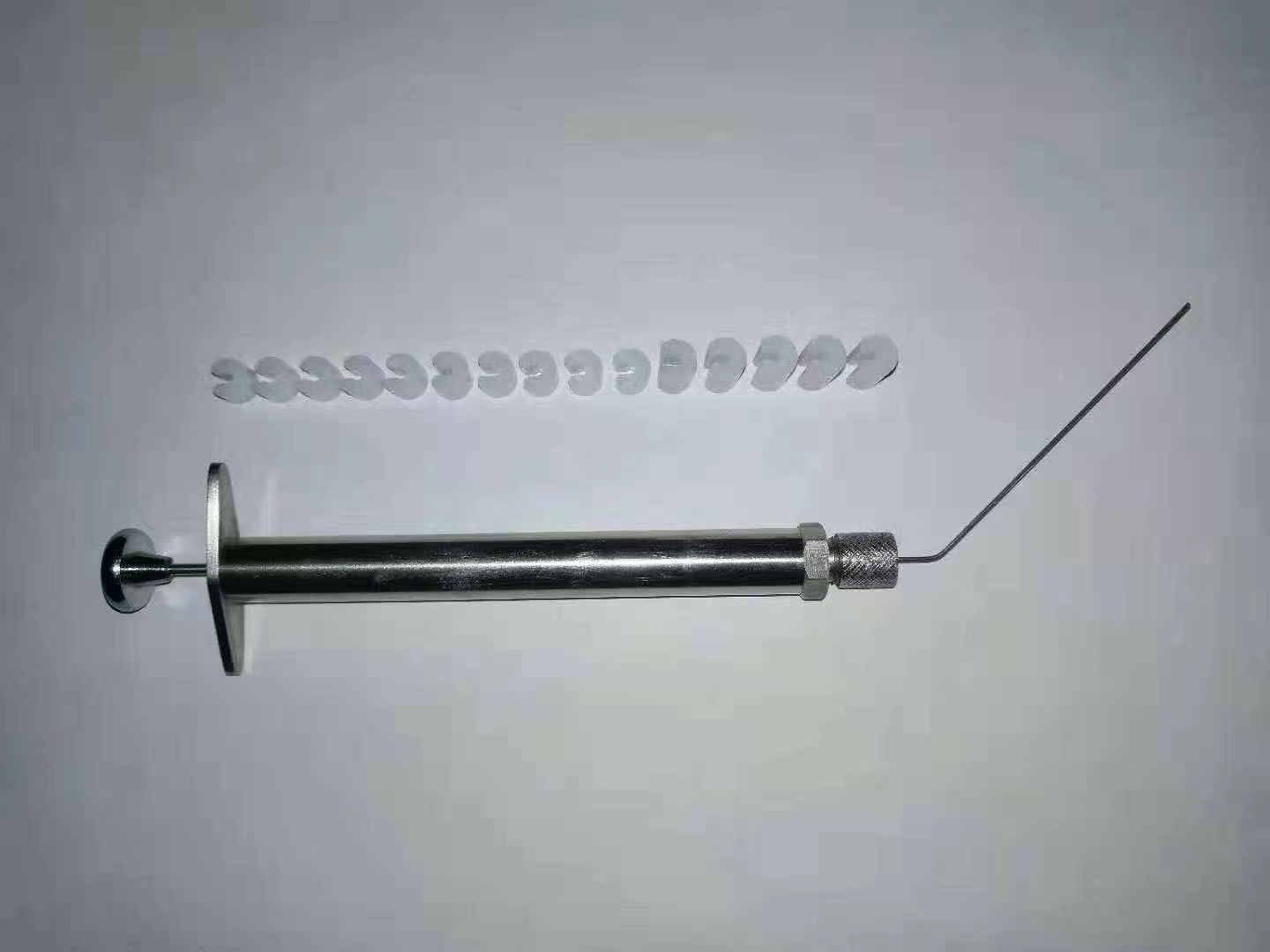
性能特点:
精确定量
较气管内滴入在各肺叶中分布更均匀
直达肺部、易于操作
更安全的提供高浓度
可输送液体、干粉样品
应用范围:
广泛应用于呼吸系统疾病、毒理学、药理学、吸入免疫、生物安全、大气污染物、化学物质毒性鉴定、药物开发与安全性评价、环境与健康等领域
风险提示:丁香通仅作为第三方平台,为商家信息发布提供平台空间。用户咨询产品时请注意保护个人信息及财产安全,合理判断,谨慎选购商品,商家和用户对交易行为负责。对于医疗器械类产品,请先查证核实企业经营资质和医疗器械产品注册证情况。
- 作者
- 内容
- 询问日期
 文献和实验
文献和实验Treating Multiorgan Fibrosis
Qiang Long, Zehua Liu, Qianwen Shao, Hongpeng Shi, Shixing Huang, Chenyu Jiang,
Bei Qian, Yiming Zhong, Xiaojun He, Xiaogang Xiang, Yang Yang, Bing Li, Xiaoxiang Yan,
Qiang Zhao,* Xiaoli Wei,* Hélder A. Santos,* and Xiaofeng Ye*
Fibrotic diseases remain a substantial health burden with few therapeutic
approaches. A hallmark of fibrosis is the aberrant activation and accumulation
of myofibroblasts, which is caused by excessive profibrotic cytokines.
Conventional anticytokine therapies fail to undergo clinical trials, as simply
blocking a single or several antifibrotic cytokines cannot abrogate the
profibrotic microenvironment. Here, biomimetic nanoparticles based on
autologous skin fibroblasts are customized as decoys to neutralize multiple
fibroblast-targeted cytokines. By fusing the skin fibroblast membrane onto
poly(lactic-co-glycolic) acid cores, these nanoparticles, termed fibroblast
membrane-camouflaged nanoparticles (FNPs), are shown to effectively
scavenge various profibrotic cytokines, including transforming growth
factor-휷, interleukin (IL)-11, IL-13, and IL-17, thereby modulating the
profibrotic microenvironment. FNPs are sequentially prepared into multiple
formulations for different administration routines. As a proof-of-concept, in
three independent animal models with various organ fibrosis (lung fibrosis,
liver fibrosis, and heart fibrosis), FNPs effectively reduce the accumulation of
myofibroblasts, and the formation of fibrotic tissue, concomitantly restoring
organ function and indicating that FNPs are a potential broad-spectrum
therapy for fibrosis management.
Q. Long, H. Shi, S. Huang, C. Jiang, B. Qian, Y. Zhong, X. He, Q. Zhao,
X. Ye
Department of Cardiovascular Surgery
Ruijin Hospital
Shanghai Jiao Tong University School of Medicine
Shanghai 200025, China
E-mail: zq11607@rjh.com.cn; yxf11612@rjh.com.cn
Z. Liu, H. A. Santos
Department of Biomedical Engineering, W.J. Kolff Institute for
Biomedical Engineering and Materials Science
University Medical Center Groningen/University of Groningen
Ant. Deusinglaan 1, Groningen 9713 AV, The Netherlands
E-mail: h.a.santos@umcg.nl
The ORCID identification number(s) for the author(s) of this article
can be found under https://doi.org/10.1002/advs.202200856
© 2022 The Authors. Advanced Science published by Wiley-VCH GmbH.
This is an open access article under the terms of the Creative Commons
Attribution License, which permits use, distribution and reproduction in
any medium, provided the original work is properly cited.
DOI: 10.1002/advs.202200856
1. Introduction
Fibrosis, or disordered fibrotic tissue formation, is characterized by the abnormal
fibroblast activation that induces excessive extracellular matrix (ECM) remodeling
and primarily accounts for multiple organ
dysfunctions.[1] The pervasive occurrence
of fibrosis in almost all diseases generates
a large healthcare burden worldwide. However, the clinical benefits of antifibrotic therapy through small molecules, such as pirfenidone and nintedanib, are usually offset
by their modest therapeutic efficacy, limited
indications and severe side effects.[2] Therefore, alternative clinical intervention modalities to target fibrosis are urgently needed.
Considering the central role of myofibroblast activation and proliferation in
fibrosis establishment,[3] recent breakthroughs have focused on the ablation
of progressive myofibroblast activation
through autologous cell-based therapy.
For example, autologous chimeric antigen
Z. Liu, H. A. Santos
Drug Research Program
Division of Pharmaceutical Chemistry and Technology
Faculty of Pharmacy
University of Helsinki
Helsinki FI-00014, Finland
Q. Shao, X. Wei
Department of Pharmacology
School of Basic Medical Sciences
Fudan University
Shanghai 200032, China
E-mail: xlwei@fudan.edu.cn
X. Xiang
Department of Infectious Diseases
Ruijin Hospital
Shanghai Jiao Tong University School of Medicine
Shanghai 200025, China
Y. Yang
Department of Thoracic Surgery
Shanghai Pulmonary Hospital
School of Medicine
Tongji University
Shanghai 200000, China
Adv. Sci. 2022, 9, 2200856 2200856 (1 of 14) © 2022 The Authors. Advanced Science published by Wiley-VCH GmbH
。一般注射部位为胸、腹或股淋巴囊。由于其皮肤很薄缺乏弹性,注射后药物易从针孔溢出,所以胸部淋巴囊注射时应将针头插入口腔,由口腔底部穿过下颌肌层进入淋巴囊,将药物注入。 (七)其它途径给药 1、呼吸道给药 呈粉尘、气体及蒸气或雾等症状存在药物或毒气,均需要通过动物呼吸道给药。如一般实验时给动物乙醚作吸入麻醉,给动物吸一定量的氨气、二氧化碳等观察呼吸、循环等变化;给动物定期吸入一定量的SO2。锯末烟雾等可造成慢性气管炎动物模型等;特别在毒物学实验中应用更为广泛。 2、皮肤给药 为了鉴定药物
抗原|抗体技术 【求助】pcgene软件 【讨论】为什么现在采的腹水呈现乳白色胶胨状? 【求助】间接ELISA的可行性问题 【讨论】DEAE-纤维素纯化抗体时PBS洗脱的问题 【交流】谁需要订购PAX5抗体的请和我联系 【求助】多克隆抗体的特异性 【求助】请问谁有大鼠Apelin和APJ抗体? 【求助】用哪种方法测兔的细胞因子? 【求助】抗血清效价测定时阴性血清吸光值 【讨论】IgG蛋白浓度用BCA定量不准么? 【求助】蛋白
1、乙醚乙醚吸入法是最常用的麻醉方法,各种动物都可应用。其麻醉量和致死量相差大,所以其安全度大。但由于乙醚局部刺激作用大,可刺激上呼吸道粘液分泌增加;通过神经反射还可扰乱呼吸、血压和心脏的活动,并且容易引起窒息,在麻醉过程中要注意。但总起来说乙醚麻醉的优点多,如麻醉深度易于掌握,比较安全,而且麻醉后恢复比较快。其缺点是需要专人负责管理麻醉,在麻醉初期出现强烈的兴奋现象,对呼吸道又有较强的刺激作用,因此,需在麻醉前给予一定量的吗啡和阿托品(基础麻醉),通常在麻醉前20~30分钟,皮下注射盐
 技术资料
技术资料暂无技术资料 索取技术资料

