

- ¥8467.50
- Lifeline Cell Technology
- 美国
- FC-0007
- 2026年02月25日

企业认证
相关产品推荐更多 >
万千商家帮你免费找货
0 人在求购买到急需产品
- 详细信息
- 文献和实验
- 技术资料
- 英文名:
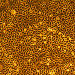
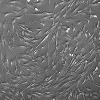
Epidermal Keratinocytes — Neonatal, Primary
- 细胞类型:
人正常原代细胞
- 物种来源:
人源
- 器官来源:
人源
- 运输方式:
干冰或液氮
- 年限:
液氮储存10年以上
- 生长状态:
冻存
- 规格:
500,000 cells/vial
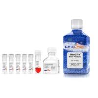
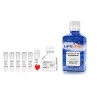
相关产品: 培养试剂 DermaLife K培养基
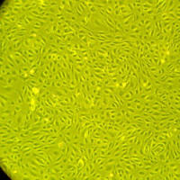
Lifeline® Normal Human Epidermal Keratinocytes neonatal (HEKn), when grown in Lifeline DermaLife K Medium, provides an ideal serum-free culture model, for the study of wound healing, toxicology or epithelial biology.
Lifeline Epidermal Keratinocytes are cryopreserved as primary cells to ensure the highest viability and plating efficiency. Cells are isolated from neonatal human foreskin and expanded once in culture vessels before cryopreservation.
- Our Neonatal Epidermal Keratinocytes are quality tested in DermaLife K Medium to ensure optimal serum-free growth over a period of at least 15 population doublings at rates equal to or greater than serum-supplemented medium.
- Keratinocytes can be grown without serum, phenol red or antimicrobials when cultured in DermaLife medium.
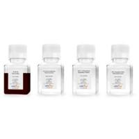
Lifeline® Epidermal Keratinocytes need not be exposed to antimicrobials or phenol red when cultured in DermaLife medium, an advantage since these supplements can cause cell stress and “masking effects” that may negatively impact experimental results. Lifeline® offers these traditional supplements, but they are not needed or recommended to achieve optimal cell performance in most research applications.

Quality Testing for Guaranteed Consistency and Reproducible Results
Lifeline® Cell Technology manufactures products using the highest quality raw materials and incorporates extensive quality assurance in every production run. Exacting standards and production procedures ensure consistent performance.

500,000 cells per vial
风险提示:丁香通仅作为第三方平台,为商家信息发布提供平台空间。用户咨询产品时请注意保护个人信息及财产安全,合理判断,谨慎选购商品,商家和用户对交易行为负责。对于医疗器械类产品,请先查证核实企业经营资质和医疗器械产品注册证情况。
 文献和实验
文献和实验利用 Millicell 培养小室培养皮肤及肺类器官的实验方案
培养基,并去除细胞培养小室膜下方的气泡。 3D 皮肤培养 10 天后,应产生大约 8 至 10 层的活性上皮层。 结果 体外皮肤类器官 (A) 新生儿皮肤组织 (B) (C) (D) 图 2. 皮肤类器官模型具有与新生儿皮肤组织相当的分层上皮形态。 H&E 染色的含有真皮和表皮分层层的体外皮肤类器官和离体皮肤切片(A、B)。丝聚蛋白染色可识别角化角质形成细胞层(棕色)(C、D)。 A B C 图 3. 皮肤类器官模型包含活跃增殖的角质形成细胞。BrdU(溴脱氧尿苷/5-溴
Delta-delta Ct method and PCR efficiencies-Real-Time PCR
we use HNEK cells...human neonatal epidermal keratinocytes. each lot was pooled from three separate neonatal donors. they usually try to get at least two ethnic groups involved, so the samples are really pretty varied
PRIMARY MOUSE KERATINOCYTE CULTURES Isolation of epidermal keratinocytes from neonatal mice is based on the protocol of Dlugosz et al., Methods Enzymol . 254:3-20 (1995). The epidermis from a newborn mouse should yield


