

- ¥2980
- LSM Bio
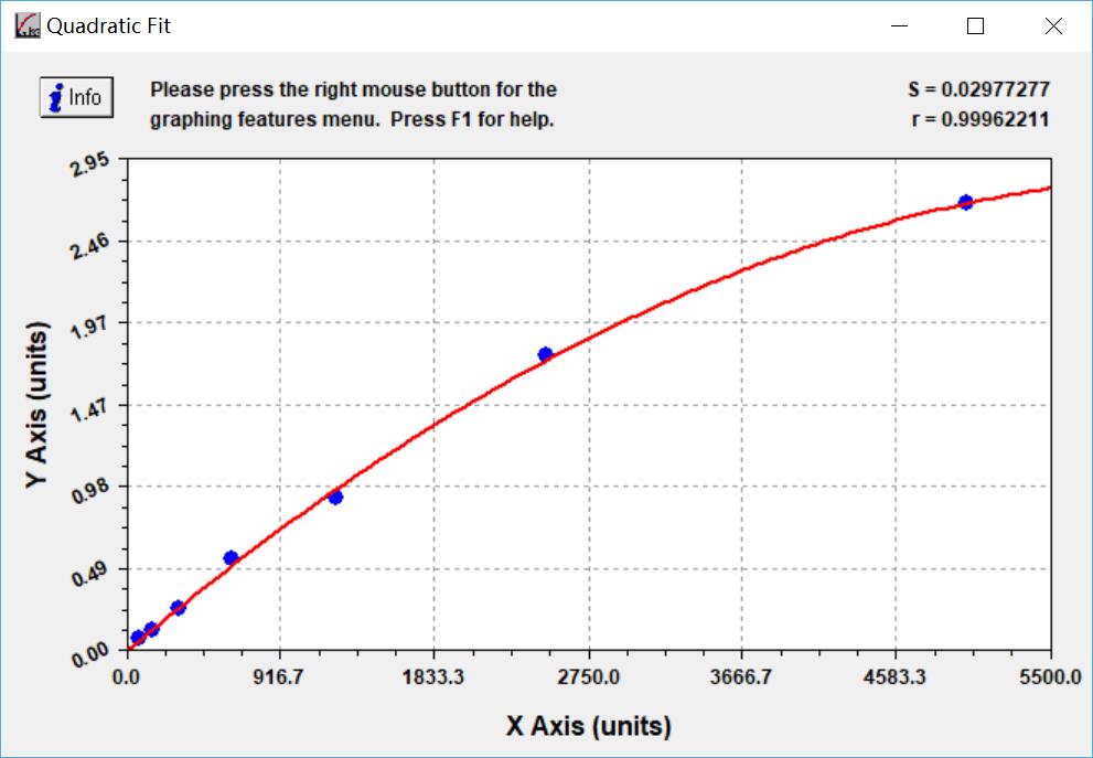
- 78-5000pg/ml
- 46.9pg/ml
- ELI-32610m
- 2026年03月10日

企业认证
相关产品推荐更多 >

Mouse Zwint ELISA Kit
¥2980
Mouse Ankk1 ELISA KIT
¥2980
Chek1 ELISA Kit| Mouse Serine/threonine-protein kinase Chk1 ELIChek1 ELISA试剂盒| Mouse Serine/threonine-protein kinase Chk1 ELI
¥2980
Mouse Peptidyl-prolyl cis-trans isomerase C, Ppic ELISA KIT
¥2980
Mouse F-box LRR-repeat protein 15, Fbxl15 ELISA KIT
¥2980
万千商家帮你免费找货
0 人在求购买到急需产品
- 详细信息
- 文献和实验
- 技术资料
- 灵敏度:
46.9pg/ml
- 库存:
50
- 供应商:
LSM Bio
- 检测范围:
78-5000pg/ml
- 检测方法:
夹心法ELISA或竞争法ELISA
- 应用:
检测 小鼠样本Protein FAM19A5, Fam19a5蛋白浓度
- 适应物种:
小鼠
- 标记物:
Protein FAM19A5, Fam19a5
- 样本:
小鼠血清,血浆,组织匀浆
- 规格:
96T
Mouse FAM19A5 ELISA KIT
Packing 96Tests
FOR RESEARCH USE ONLY; NOT FOR THERAPEUTIC OR DIAGNOSTIC APPLICATIONS! PLEASE READ ENTIRE PROCEDURE!
Gene Name FAM19A5
Protein Name protein FAM19A5
Alternative Name
FAM19A5,protein FAM19A5; QLLK5208; TAFA-5; TAFA5; UNQ5208; TAFA protein 5; chemokine-like protein TAFA-5; family with sequence similarity 19 (chemokine (C-C motif)-like), member A5; family with sequence similarity 19 member A5, C-C motif chemokine lik
Intended use
Mouse FAM19A5 ELISA KIT allows for the in vitro quantitative determination of FAM19A5 concentrations in serum, plasma, tissue homogenates, cell culture supernates or other biological fluids.
|
Reagent |
Quantity |
|
Assay plate |
1 |
|
Standard |
2 |
|
Sample Diluent |
1 × 20mL |
|
Assay Diluent A |
1 × 10mL |
|
Assay Diluent B |
1 × 10mL |
|
Detection Reagent A |
1 × 120μL |
|
Detection Reagent B |
1 × 120μL |
|
Wash Buffer(25 x concentrate) |
1 × 30mL |
|
Substrate |
1 × 10mL |
|
Stop Solution |
1 × 10mL |
|
Plate sealer |
5 |
Test principle
The microtiter plate provided in Mouse FAM19A5 ELISA KIT has been pre-coated with an FAM19A5 antibody specific to FAM19A5. Standards or samples are then added to the appropriate microtiter plate wells with a biotin-conjugated antibody preparation specific for FAM19A5 and then avidin conjugated to Horseradish Peroxidase (HRP) is added to each microplate well and incubated. Then a TMB substrate solution is added to each well. Only those wells that contain FAM19A5, biotin-conjugated antibody and enzyme-conjugated Avidin will exhibit a change in color. The enzyme-substrate reaction is terminated by the addition of a sulphuric acid solution and the color change is measured spectrophotometrically at a wavelength of 450 nm ± 2 nm. The concentration of FAM19A5 in the samples is then determined by comparing the O.D. of the samples to the standard curve.
Sample collection and storage
Serum - Use a serum separator tube (SST) and allow samples to clot for 30 minutes before centrifugation for 15 minutes at approximately 1000 × g. Remove serum and assay immediately or aliquot and store samples at -20鈩?/p>
or -80鈩?.
Plasma - Collect plasma using EDTA or heparin as an anticoagulant. Centrifuge samples for 15 minutes at 1000 × g at 2鈩? 8鈩?nbsp; within 30 minutes of collection. Store samples at -20鈩?nbsp; or -80鈩?.
Tissue homogenates - The preparation of tissue homogenates will vary depending upon tissue type. For this assay, tissue was rinsed with ice-cold 1×PBS to remove excess blood, homogenized in ice-cold 1×PBS and stored overnight at ≤-20鈩?. In most cases, 10% homogenate (eg.1g of tissue in 10mL of ice-cold 1×PBS) is recommended. After two freeze-thaw cycles were performed to break the cell membranes, the homogenates were centrifuged
for 5 minutes at 5000 x g. Remove the supernate and assay immediately or aliquot and store at ≤-20鈩?nbsp; .
Cell culture supernates and Other biological fluids - Remove particulates by centrifugation and assay immediately or aliquot and store samples at -20鈩?or -80鈩?.
Fresh samples are first choice. If not, avoid freeze-thaw of samples.
Reagent preparation
Standard - Please refer to the Data Sheet inserting in the ELISA kit.
Detection Reagent A and B - Dilute to the working concentration using Assay Diluent A and B (1:100), respectively.
Wash Buffer - If crystals have formed in the concentrate, warm to room temperature and mix gently until the crystals have completely dissolved. Dilute 30mL of Wash Buffer Concentrate into deionized or distilled water to prepare 750 mL of Wash Buffer.
Assay procedure
Allow all reagents to reach room temperature (Please do not dissolve the reagents at 37鈩?directly). All the reagents should be mixed thoroughly by gently swirling before pipetting. Avoid foaming. Keep appropriate numbers
of strips for 1 experiment and remove extra strips from microtiter plate. Removed strips should be resealed and stored at -20鈩?until the kits expiry date. Prepare all reagents, working standards and samples as directed in the
previous sections. Please predict the concentration before assaying. If values for these are not within the range of the standard curve, users must determine the optimal sample dilutions for their particular experiments.
1. Add 100 μL of Standard, Blank, or Sample per well. Cover with the Plate sealer. Incubate for 2 hours at 37鈩?
2. Remove the liquid of each well, don’t wash. Add 100μL of Detection
Reagent A working solution to each well. Cover with the Plate sealer. Incubate for 1 hour at 37鈩? Detection Reagent A working solution may appear cloudy. Warm to room temperature and mix gently until solution
appears uniform.
3. Aspirate each well and wash, repeating the process three times for a total of three washes. Wash by filling each well with Wash Buffer (approximately 400 μL) using a squirt bottle, multi-channel pipette, manifold dispenser or autowasher, and let it sit for 1~2 minutes. Complete removal of liquid at each step is essential for good performance. After the last wash, remove any remaining Wash Buffer by aspirating or decanting. Invert the plate and blot it against clean paper towels.
4. Add 100μL of Detection Reagent B working solution to each well. Cover with a new Plate sealer. Incubate for 1 hour at 37鈩?
5. Repeat the aspiration/wash process for 5 times as conducted in step 3.
6. Add 90μL of Substrate Solution to each well. Cover with a new Plate sealer. Incubate within 15-30 minutes at 37鈩? Protect from light.
7. Add 50μL of Stop Solution to each well. If color change does not appear
uniform, gently tap the plate to ensure thorough mixing.
8. Determine the optical density of each well at once, using a microplate reader set to 450 nm.
Calculation of results
Average the duplicate readings for each standard, control, and sample and sample, then subtract the average zero standard optical density. Create a standard curve by reducing the data using computer software capable of
generating a four parameter logistic (4-PL) curve-fit. As an alternative, construct a standard curve by plotting the mean absorbance for each standard on the x-axis against the concentration on the y-axis and draw a best fit curve through the points on the graph. The data may be linearized by plotting the log of the FAM19A5 concentrations versus the log of the O.D. and the best fit line can be determined by regression analysis. It is recommended to use professional software to do this calculation, such as CurveExpert. This procedure will produce an adequate but less precise fit of the data. If samples have been diluted, the concentration read from the standard curve must be multiplied by the dilution factor.
Important note
1. Please carefully reconstitute Standards or working Detection Reagent A and B according to the instruction, and avoid foaming and mix gently until the crystals have completely dissolved. The reconstituted Standards, Detection Reagent A and B can be used only once.
2. To ensure accurate results, proper adhesion of plate sealers during incubation steps is necessary. Do not allow wells to sit uncovered for extended periods between incubation steps. Once reagents have been added to the well strips, DO NOT let the strips DRY at any time during the assay.
3. To avoid cross-contamination, change pipette tips between additions of each standard level, between sample additions, and between reagent additions. Also, use separate reservoirs for each reagent.
4. The wash procedure is critical. Insufficient washing will result in poor precision and falsely elevated absorbance readings.
5. Substrate Solution is easily contaminated. Please protect it from light.
6. ELISA Kits from different batches may be a little different in detection range, sensitivity and color developing time.Please perform the experiment exactly according to printed instruction inside in the kit while electronic ones from our website is for reference only.
7. Do not substitute reagents from one lot to another. Use only the reagents in the same kit supplied by manufacturer.
8. Even the same operator might get different results in two separate experiments. In order to get better reproducible results, the operation of every step in the assay should be controlled. Furthermore, a preliminary experiment before assay for each batch is recommended.
9. Each ELISA kit has been strictly passed QC test. However, results from end users might be inconsistent with our in-house data due to some unexpected transportation conditions or different lab equipments. Intra-assay variance among kits from different batches might arise from above factors, too.
10. ELISA Kits from different manufacturers for the same item might produce different results, since we haven’t compared our products with other manufacturers.
11. Period of validity: six months.
Precaution
The Stop Solution provided with Mouse FAM19A5 ELISA KIT is an acid solution. Wear eye, hand, face, and clothing protection when using this material.
风险提示:丁香通仅作为第三方平台,为商家信息发布提供平台空间。用户咨询产品时请注意保护个人信息及财产安全,合理判断,谨慎选购商品,商家和用户对交易行为负责。对于医疗器械类产品,请先查证核实企业经营资质和医疗器械产品注册证情况。
 文献和实验
文献和实验Article: Quercetin and lithium chloride potentiate the protective effects of carvedilol against renal ischemia-reperfusion injury in high-fructose, high-fat diet-fed swiss albino mice independent of renal lipid signaling
Author:Asmaa M.RezkabIslam A.A.E.-H.IbrahimaMona F.MahmoudaAmr A.A.Mahmouda1
Received 6 September 2020, Revised 12 October 2020, Accepted 28 October 2020, Available online 4 November 2020.
Chemico-Biological Interactions
Available online 4 November 2020, 109307
doi: doi.org/10.1016/j.cbi.2020.109307
, incubate for 30 min at 37°C. ( 5g powdered milk in 95 ml PBS)5 Rinse with PBS 3x.6 Add 50 µl of antibody, incubate 30 min at 37°C.7 Rinse with PBS 3x.8 Add 50 µl of goat anti-mouse IgG conjugate with alkaline phosphatase.(1 µl of conjugate in 4 ml of PBS
反应发生的风险,故而在合成磷酸化多肽时,需要对氨基酸投料量、反应方法以及反应时长等进行优化调整以期达到更理想、更经济的合成效果。我们有针对性地对磷酸化多肽合成条件进行了探索和调整,采用最终的优化条件成功合成了含有多达六个磷酸化丝氨酸残基的多肽:FAM-Ahx-X(pS)XX(pS)X(pS)X(pS)XX(pS)X(pS)-NH2(客户肽,详细序列未给出;其氨基端标记FAM以进行荧光检测),经过RP-HPLC纯化后最终纯品的纯度高达95%(见Figure 3)。 参考文献: 1. P
The indirect ELISA is used primarily to determine the strength and/or amount of antibody response in a sample, whether it is from the serum of an immunized animal or the cell supernatant from growing hybridoma clones. Procedure 1. All incubations
 技术资料
技术资料
