


- ¥1700 - 4000
- GeneTex

- 美国
- GTX101691
- 2025年07月16日
- WB, IHC-P, IHC-Wm, IP, IHC
- Rabbit
- Human, Mouse, Rat, Zebrafish

企业认证
相关产品推荐更多 >

![UNC13B antibody [N2N3]](https://img1.dxycdn.com/2023/0516/045/6925717576889828561-14.jpg!wh200)

万千商家帮你免费找货
0 人在求购买到急需产品
- 详细信息
- 文献和实验
- 技术资料
- 免疫原:
Recombinant protein encompassing a sequence within the center region of human ILK. The exact sequence is proprietary.
- 亚型:
IgG
- 形态:
Liquid
- 保存条件:
Store as concentrated solution. Centrifuge briefly prior to opening vial. For short-term storage (1-2 weeks), store at 4ºC. For long-term storage, aliquot and store at -20ºC or below. Avoid multiple freeze-thaw cycles.
- 克隆性:
Polyclonal
- 标记物:
Unconjugated
- 适应物种:
Human, Mouse, Rat, Zebrafish
- 保质期:
12 months from the shipping date of the product.
- 抗原来源:
Human
- 目录编号:
GTX101691
- 级别:
Primary Antibodies
- 库存:
Available
- 供应商:
GeneTex
- 宿主:
Rabbit
- 应用范围:
WB, IHC-P, IHC-Wm, IP, IHC
- 浓度:
0.8 mg/ml (Please refer to the vial label for the specific concentration.)
- 靶点:
ILK
- 抗体英文名:
ILK antibody [N1C1]
- 抗体名:
ILK 抗体 [N1C1]
- 规格:
100 μl/25 μl
| 规格: | 100 μl | 产品价格: | ¥4000.0 |
|---|---|---|---|
| 规格: | 25 μl | 产品价格: | ¥1700.0 |
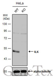
Wild-type (WT) and ILK knockout (KO) HeLa cell extracts (30 μg) were separated by 10% SDS-PAGE, and the membrane was blotted with ILK antibody [N1C1] (GTX101691) diluted at 1:500. The HRP-conjugated anti-rabbit IgG antibody (GTX213110-01) was used to detect the primary antibody.

Whole cell extract (30 μg) was separated by 10% SDS-PAGE, and the membrane was blotted with ILK antibody [N1C1] (GTX101691) diluted at 1:500.
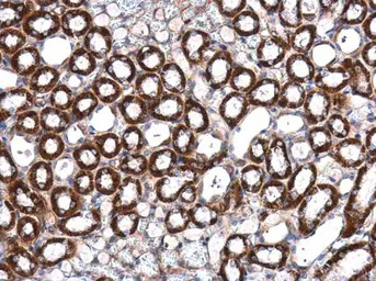
ILK antibody [N1C1] detects ILK protein at cytoplasm in mouse kidney by immunohistochemical analysis.
Sample: Paraffin-embedded mouse kidney.
ILK antibody [N1C1] (GTX101691) diluted at 1:500.
Antigen Retrieval: Citrate buffer, pH 6.0, 15 min
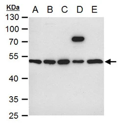
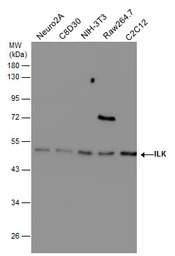
ILK antibody [N1C1] detects ILK protein by western blot analysis.
A. 30 μg Neuro2A whole cell extract
B. 30 μg C8D30 whole cell extract
C. 30 μg NIH-3T3 whole cell extract
D. 30 μg Raw 264.7 whole cell extract
E. 30 μg C2Cl2 whole cell extract
10 % SDS-PAGE
ILK antibody [N1C1] (GTX101691) dilution: 1:1000

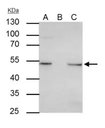
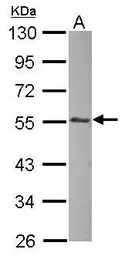
Sample(30 μg of whole cell lysate)
A:A431(GTX27909)
7.5% SDS PAGE
GTX101691 diluted at 1:500
ILK antibody [N1C1] immunoprecipitates ILK protein in IP experiments.
IP samples: HeLa whole cell extract
A. 30 μg HeLa whole cell extract
B. Control with 4 μg of preimmune Rabbit IgG
C. Immunoprecipitation of ILK protein by 4 μg ILK antibody [N1C1] (GTX101691)
10 % SDS-PAGE
The immunoprecipitated ILK protein was detected by ILK antibody [N1C1] (GTX101691) diluted at 1:500.
[EasyBlot anti-rabbit IgG (GTX221666-01) was used as a secondary reagent]
Various whole cell extracts (30 μg) were separated by 10% SDS-PAGE, and the membrane was blotted with ILK antibody [N1C1] (GTX101691) diluted at 1:500.
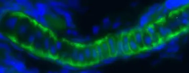
Immunofluorescent image of a pharyngeal cartilage section of a zebrafish embryo using ILK antibody [N1C1] (GTX101691) at a 1:200 dilution. ILK (Green)(This image was provided courtesy of the Schilling Lab at UC, Irvine.)
Sample (30 μg of whole cell lysate)
A: zebrafish muscle
10% SDS PAGE
GTX101691 diluted at 1:1000

Immunohistochemical analysis of paraffin-embedded zebrafish tissue, using ILK antibody [N1C1] (GTX101691) at 1:300 dilution.

Immunohistochemical analysis of agarose-embedded zebrafish embryo, using ILK antibody [N1C1] (GTX101691) at 1:100 dilution. (This image was provided courtesy of the Schilling Lab at UC, Irvine.)
风险提示:丁香通仅作为第三方平台,为商家信息发布提供平台空间。用户咨询产品时请注意保护个人信息及财产安全,合理判断,谨慎选购商品,商家和用户对交易行为负责。对于医疗器械类产品,请先查证核实企业经营资质和医疗器械产品注册证情况。
 文献和实验
文献和实验Chen YL et al., Sci Rep 2021 (PMID:33436772)
Wantoch von Rekowski K et al., Biomolecules 2019 (PMID:31779287)
Yen YC et al., Oncotarget 2015 (PMID:26540630)
Huang CY et al., Mol Neurobiol 2014 (PMID:24771044)
[资源] 所有的看家基因(housekeeping genes)列表+引物设计服务
phosphatase 2 (formerly 2A), catalytic subunit, beta isoform (PPP2CB), mRNA 1195 NM_004910 Homo sapiens phosphatidylinositol transfer protein, membrane-associated (PITPNM), mRNA 869 NM_004517 Homo sapiens integrin-linked kinase (ILK), mRNA 654 NM
 技术资料
技术资料暂无技术资料 索取技术资料

