

- ¥1700 - 4000
- GeneTex

- 美国
- GTX113016
- 2025年07月16日
- WB, ICC/IF, IHC-P, IHC-Fr
- Rabbit
- Human, Mouse, Rat, Zebrafish, Vole, Calf

企业认证
相关产品推荐更多 >

![FRK antibody [5F3]](https://img1.dxycdn.com/2023/0711/451/6106825198874858761-14.jpg!wh200)

![BPGM antibody [N1C3]](https://img1.dxycdn.com/2023/0516/895/8887749495766728561-14.jpg!wh200)
万千商家帮你免费找货
0 人在求购买到急需产品
- 详细信息
- 文献和实验
- 技术资料
- 免疫原:
Recombinant protein encompassing a sequence within the center region of human Tyrosine Hydroxylase. The exact sequence is proprietary.
- 亚型:
IgG
- 形态:
Liquid
- 保存条件:
Store as concentrated solution. Centrifuge briefly prior to opening vial. For short-term storage (1-2 weeks), store at 4ºC. For long-term storage, aliquot and store at -20ºC or below. Avoid multiple freeze-thaw cycles.
- 克隆性:
Polyclonal
- 标记物:
Unconjugated
- 适应物种:
Human, Mouse, Rat, Zebrafish, Vole, Calf
- 保质期:
12 months from the shipping date of the product.
- 抗原来源:
Human
- 目录编号:
GTX113016
- 级别:
Primary Antibodies
- 库存:
Available
- 供应商:
GeneTex
- 宿主:
Rabbit
- 应用范围:
WB, ICC/IF, IHC-P, IHC-Fr
- 浓度:
0.21 mg/ml (Please refer to the vial label for the specific concentration.)
- 靶点:
Tyrosine Hydroxylase
- 抗体英文名:
Tyrosine Hydroxylase antibody
- 抗体名:
Tyrosine Hydroxylase 抗体
- 规格:
100 μl/25 μl
| 规格: | 100 μl | 产品价格: | ¥4000.0 |
|---|---|---|---|
| 规格: | 25 μl | 产品价格: | ¥1700.0 |
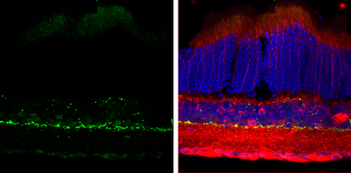
Tyrosine Hydroxylase antibody detects Tyrosine Hydroxylase protein by immunohistochemical analysis.
Sample: Frozen sectioned adult mouse retina.
Green: Tyrosine Hydroxylase protein stained by Tyrosine Hydroxylase antibody (GTX113016) diluted at 1:250.
Red: beta Tubulin 3/ TUJ1, stained by beta Tubulin 3/ TUJ1 antibody [GT11710] (GTX631836) diluted at 1:250.
Blue: Fluoroshield with DAPI (GTX30920).
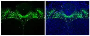
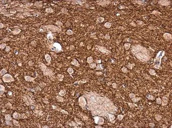
Tyrosine Hydroxylase antibody detects Tyrosine Hydroxylase protein in midbrain dopaminergic neurons by immunohistochemical analysis.
Sample: Paraffin-embedded mouse brain.
Green: Tyrosine Hydroxylase stained by Tyrosine Hydroxylase antibody (GTX113016) diluted at 1:1000.
Blue: Fluoroshield with DAPI (GTX30920).
Antigen Retrieval: Citrate buffer, pH 6.0, 15 min
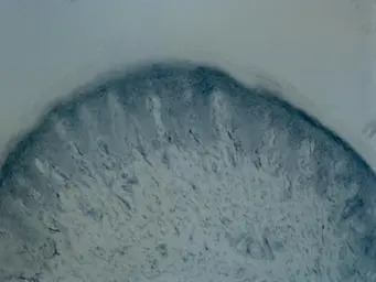
Immunohistochemical analysis of Rat hindlimb pad skin tissue (PFA-fixed frozen sections), using Tyrosine Hydroxylase(GTX113016) antibody at 1:100 dilution.
Antigen Retrieval: Citrate buffer, pH 6.0, 15 min
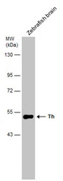
Zebrafish tissue extract (30 μg) was separated by 7.5% SDS-PAGE, and the membrane was blotted with Tyrosine Hydroxylase antibody (GTX113016) diluted at 1:500.
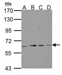
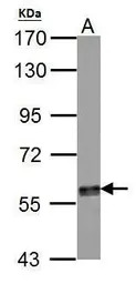
Tyrosine Hydroxylase antibody detects TH protein by western blot analysis.
A. 30 μg NT2D1 whole cell lysate/extract
B. 30 μg PC-3 whole cell lysate/extract
C. 30 μg U87-MG whole cell lysate/extract
D. 30 μg SK-N-SH whole cell lysate/extract
7.5% SDS-PAGE
Tyrosine Hydroxylase antibody (GTX113016) dilution: 1:500
The HRP-conjugated anti-rabbit IgG antibody (GTX213110-01) was used to detect the primary antibody.
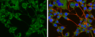
Tyrosine Hydroxylase antibody detects Tyrosine Hydroxylase protein at cytoplasm by immunofluorescent analysis.
Sample: U-87 MG cells were fixed in 4% PFA at RT for 15 min.
Green: Tyrosine Hydroxylase protein stained by Tyrosine Hydroxylase antibody (GTX113016) diluted at 1:400.
Red: beta Tubulin 3/ TUJ1 protein stained by beta Tubulin 3/ TUJ1 antibody (GTX631836) diluted at 1:200.
Blue: Hoechst 33342 staining.
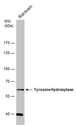
Rat tissue extract (50 μg) was separated by 7.5% SDS-PAGE, and the membrane was blotted with Tyrosine Hydroxylase antibody (GTX113016) diluted at 1:1000.
Tyrosine Hydroxylase antibody detects TH protein by western blot analysis.
A. 50 μg mouse brain lysate/extract
7.5% SDS-PAGE
Tyrosine Hydroxylase antibody (GTX113016) dilution: 1:500
The HRP-conjugated anti-rabbit IgG antibody (GTX213110-01) was used to detect the primary antibody.
Tyrosine Hydroxylase antibody detects Tyrosine Hydroxylase protein at cytoplasm in rat brain by immunohistochemical analysis.
Sample: Paraffin-embedded rat brain.
Tyrosine Hydroxylase antibody (GTX113016) diluted at 1:2500.
Antigen Retrieval: Citrate buffer, pH 6.0, 15 min
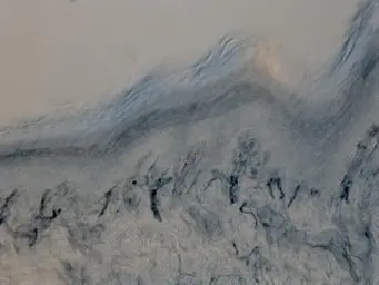
Immunohistochemical analysis of Rat hindlimb pad skin tissue (PFA-fixed frozen sections), using Tyrosine Hydroxylase(GTX113016) antibody at 1:100 dilution.
Antigen Retrieval: Citrate buffer, pH 6.0, 15 min
风险提示:丁香通仅作为第三方平台,为商家信息发布提供平台空间。用户咨询产品时请注意保护个人信息及财产安全,合理判断,谨慎选购商品,商家和用户对交易行为负责。对于医疗器械类产品,请先查证核实企业经营资质和医疗器械产品注册证情况。
 文献和实验
文献和实验Wu CY et al., Brain Stimul 2020 (PMID:32289709)
Santana Y et al., J Clin Med 2019 (PMID:30678046)
Li Y et al., Oxidative Medicine and Cellular Longevity 2018 (Epub)
Park CH et al., J Vet Med Sci 2018 (PMID:29526867)
Thomas MG et al., Cell Death Dis 2013 (PMID:23764850)
Zhang L et al., EMBO Mol Med 2017 (PMID:28818835)
Xiao JJ et al., Oxid Med Cell Longev 2015 (PMID:26146528)
Arai A et al., Behav Brain Res 2016 (PMID:27522019)
Ma C et al., Life Sci 2016 (PMID:26612350)
Yamamoto S et al., Front Immunol 2022 (PMID:35911740)
Wejdane El Manaa et al., Autophagy 2021 (PMID:34030589)
Lasse Reimer et al., Sci Rep 2022 (PMID:35260646)
Song Y et al., Neurosci Bull 2021 (PMID:34954810)
Di Nisio A et al., Environ Int 2022 (PMID:34781208)
Fede C et al., Sci Rep 2021 (PMID:34135423)
Wu J et al., Oxid Med Cell Longev 2020 (PMID:32724496)
Kawabe M et al., Neuropeptides 2021 (PMID:33721592)
Li Y et al., Oxid Med Cell Longev 2018 (PMID:30046373)
Phenylalanine Hydroxylase, Tyrosine Hydroxylase, and Tryptophan Hydroxylase
There are four enzymes that utilize reduced pteridine as an electron donor and incorporate one atom of oxygen into their substrates. They are phenylalanine 4-monooxygenase (phenylalanine hydroxylase, PAH, EC 1.14.16.l), tyrosine 3-monooxygenase
Quantification of Tyrosine Hydroxylase mRNA
The main biochemical characteristic of Parkinson’s disease (PD) is reduction of the neurotransmitter dopamine and the dopamine-synthesizing enzyme system, including tyrosine hydroxylase (TH, tyrosine 3-monooxygenase, EC 1.14.16.2
Proteomic Method for Identification of Tyrosine-Nitrated Proteins
Biologic nitration of protein tyrosine (to form 3-nitrotyrosine) is a recently described phenomenon that is associated with many diseases. We have devised a proteomic methodology to identify these modified proteins. This utilizes protein
 技术资料
技术资料暂无技术资料 索取技术资料

