


- ¥1500
- ATCC、DSMZ、ECACC、RIKEN
- 江苏
- CL1291
- 2026年06月21日

企业认证
相关产品推荐更多 >


万千商家帮你免费找货
0 人在求购买到急需产品
- 详细信息
- 文献和实验
- 技术资料
- 英文名:
SV40 MES 13
- 库存:
100万
- 供应商:
欣润生物
- 肿瘤类型:
否
- 细胞类型:
细胞系
- ATCC Number:
CL1291
- 品系:
小鼠
- 组织来源:
详见说明
- 相关疾病:
详见说明
- 物种来源:
小鼠
- 免疫类型:
不详
- 细胞形态:
多角形
- 是否是肿瘤细胞:
否
- 器官来源:
详见说明
- 运输方式:
新鲜或干冰
- 年限:
成年
- 生长状态:
贴壁生长
- 规格:
T25方瓶
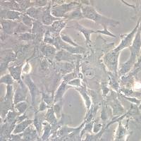
- 细胞名称:SV40 MES 13细胞(小鼠肾小球系膜细胞)
- 形态:多角型,贴壁生长
- 含量:>1x10⁶ 个/瓶
- 污染:支原体、细菌、酵母和真菌检测为阴性
- 规格:T25瓶或者1mL冻存管包装
二、细胞接收后的处理:
1、贴壁细胞
- 收到T25方瓶细胞后,请检查是否漏液,如果漏液,请拍照片发给我们(冻存管细胞收到后直接37℃水浴复苏或直接放置于液氮中长期储存)。
- 请先在显微镜下确认细胞生长状态,去掉封口膜并将T25瓶置于37℃培养约2-3h。
- 弃去T25瓶中的培养基,换用新鲜的完全培养基。
- 如果细胞长满(90%以上)请及时进行细胞传代。
- 接到细胞次日,请检查细胞是否污染,若发现污染或疑似污染,请及时与我们取得联系。
2、悬浮细胞
- 收到细胞后,请检查是否漏液,如果漏液,请拍照片发给我们。
- 请先在显微镜下确认细胞生长状态,去掉封口膜并将15ml离心管置于37℃培养约2-3h。
- 1200rpm离心5min,弃去15ml离心管中的培养基,细胞沉淀用新鲜的完全培养基重悬并培养。
- 如果细胞长满(90%以上)请及时进行细胞传代。
- 接到细胞次日,请检查细胞是否污染,若发现污染或疑似污染,请及时与我们取得联系。
本公司的细胞培养操作规程,供参考
一、培养基及培养冻存条件准备:
- 准备DMEM/F12培养基,90%;优质胎牛血清,10%。
- 培养条件: 气相:空气,95%;二氧化碳,5%。 温度:37℃,培养箱湿度为70%-80%。
- 冻存液:90%血清,10%DMSO,现用现配。液氮储存。
对于贴壁细胞,传代可参考以下方法:
- 弃去培养上清,用不含钙、镁离子的PBS润洗细胞1-2次。
- 加2ml消化液(0.25%Trypsin-0.53mM EDTA)于培养瓶中,置于37℃培养箱中消化2-3分钟,然后在显微镜下观察细胞消化情况,若细胞大部分变圆并脱落,迅速拿回操作台,轻敲几下培养瓶后加入3ml此细胞的培养基终止消化。
- 轻轻吹打后吸出,移入15ml离心管中,在1200RPM条件下离心5分钟,弃去上清液,加入1mL培养液后吹匀。
- 移入到事先准备好的含有5ml培养基的T-25培养瓶中或含有14ml培养基的T-75培养瓶中培养。
3)细胞冻存:待细胞生长状态良好时,可进行细胞冻存。贴壁细胞冻存时,先要消化处理并进行细胞计数。消化方法按照细胞传代方法的1-3步骤进行,最后的重悬液使用血清。悬浮细胞直接计数后离心,用血清重悬浮,加DMSO至最终浓度为10%。加入DMSO后迅速混匀,按每1ml的数量分配到冻存管中。本公司按每个冻存管细胞数目大于1X10⁶个细胞冻存。
注意事项:
1. 收到冻存管细胞后,若发现干冰已挥发干净、冻存管瓶盖脱落、破损及细胞有污染,请立即与我们联系。
2. 所有动物细胞均视为有潜在的生物危害性,必须在二级生物安全台内操作,并请注意防护,所有废液及接触过此细胞的器皿需要灭菌后方能丢弃。
3. 细胞用途:仅供科研使用。
发货方式:
复苏后发货:我们复苏细胞后发货,货期一周左右,免运费。(气温较好建议复苏后发货)
冻存发货(干冰运输):需额外增加干冰运费,选择干冰运输的我们发两管细胞,为了保证客户接种可靠性多发一管。(气温低于0℃须冻存发货)
细胞发货采取专业的运输包装,并选择最快捷的运输方式(顺丰速运或其他空运快递)
Molecular cloning of a ligand for the EPH-related receptor protein-tyrosine kinase Htk
Hemoglobin Is Expressed by Mesangial Cells and Reduces Oxidant Stress
Hemoglobin (Hb) serves as the main oxygen transporter in erythrocytes, but it is also expressed in nonhematopoietic organs, where it serves an unknown function. In this study, microarray and proteomic analyses demonstrated Hb expression in the kidney. Rat kidneys were perfused extensively with saline, and glomeruli were isolated by several techniques (sieving, manual dissection, and laser capture-microdissection). Reverse transcriptase–PCR revealed glomerular α- and β-globin expression, and immunoblotting demonstrated expression of the protein. hybridization studies showed expression of the globin subunits in the mesangium, and immunostaining confirmed this localization of Hb. Furthermore, globin mRNA expression was detected in primary cultures of rat mesangial cells but not in cultured glomerular endothelial or epithelial cells. For investigation of Hb function in mesangial cells, the SV40-MES13 murine mesangial cell line was transfected with a vector expressing α- and β-globins; this overexpression reduced production of hydrogen peroxide–induced intracellular radical oxygen species and enhanced cell viability against oxidative stress. In summary, Hb is expressed by rat mesangial cells, and its potential functions may include antioxidative defense.
风险提示:丁香通仅作为第三方平台,为商家信息发布提供平台空间。用户咨询产品时请注意保护个人信息及财产安全,合理判断,谨慎选购商品,商家和用户对交易行为负责。对于医疗器械类产品,请先查证核实企业经营资质和医疗器械产品注册证情况。
 文献和实验
文献和实验Chicken intestinal epithelial cells were obtained from NEWGAINBIO company. Cells were cultured on 37℃, with 5% CO2, in the Ham’s F-12 Nutrient (DMEM/12) that contained the following supplementations: fetal bovine serum (5%), in-sulin (5 µg/mL), transferrin (5 µg/mL), selenium (5 ng/mL), epidermal growth factor (5 ng/mL) and penicillin-streptomycin (100–100 U/mL) for cell culturing (full DMEM/12). Experiments were performed with chicken intestinal epithelial cells and working solutions were prepared with plain DMEM/12 without supplementation. For the investigations, cells were seeded onto 96-well, 24-well or 6-well polystyrene cell culture plates.
Primary hVICs (passage 2) were cultured to 50–60% confluence and infected with pGMLV-SV40T-puro lentivirus (NewgainBio, Wuxi, China) at a multiplicity of infection of 80 supplemented with 5 µg/mL polybrene (Sigma-Aldrich, Buchs, Switzerland).
Tissue was cultured until cells became visible around the tissue, and when the fusion reached 90% (FIGURE 1A) §ask ¦lled with the prepared culturing medium was sent to the company for further immortalisation. Cell immortalisation was done for cell stability and longer-term use. Immortalised cells were cultured with 10% FBS and 1% PS in the DMEM medium. After the cells multiplied and merged, they were routinely passed and grown ( NEWGAINBIO Inc. Wuxi, Jiangsu, China) (FIGURE 1B-C).
Mouse primary cultured renal vascular ECs and VSMCs were obtained from Newgainbio company, which were tested by Factor VIII and α-smooth muscle actin (α-SMA), the marker of ECs and VSMCs. RNeasy Mini Kit was used for RNA extraction, and the above protocols were repeated.
Porcine primary colon epithelial cells (Newgainbio company, Wuxi,China) were cultured in Dulbecco's Modified Eagle's Medium (Solarbio, Beijing, China) containing 10 % fetal bovine serum (BioInd, Kiryat shmona, Lsrael) at 37 ◦C and 5 % CO2 humidity.
cell Biol,1977,74(1):68-85. [3]张学明,李德雪,李子义,等.小鼠精原干细胞的形态结构特点及其细胞化学和免疫组化特性[J].中国兽医学报,1999,19(4):363~367. [4]Hofmann MC, Narisawa S, Hess RA, et al. Immortalization of germ cells and somatic testicular cells using the SV40 large T antigen[J]. Exper
)猴病毒40(SV40) 猴病毒40(SV40)的基因组常用来作为克隆载体,把外源DNA转入哺乳类细胞。猴病毒40是球形动物病毒,直径40nm,呈20面体,有一个共价闭环的双链DNA基因组,全长5244bp。它在猴肾中增殖。被SV40感染的细胞都会出现SV40的T抗原。早已证明完整的小鼠染色体β珠蛋白基因(包括所有的间插顺序和两侧顺序)整合在SV40中后,在被感染的猴肾细胞中都能正确地转录和翻译。表明猴肾细胞的拼接系统能有效地处理小鼠β珠蛋白的初级转录本。 可是,SV40作为克隆载体有其局限
摩尔)的对照报告基因酶(图3B),并且这两个酶的特异活力相似。 图2 用双荧光素酶报告基因测试由萤火虫和Renilla荧光素酶产生的发光。CHO细胞(1×10⁶ /60mm培养板)共转染pGL3Control和pRL SV40载体DNA.细胞用PBS洗涤后,加入400μl PLB制成裂解液。将20μl小量细胞裂解液和100μl荧光素酶测试试剂II(LARII)混合,萤火虫荧光素酶的活力立即用荧光照度仪检测(细线示踪)。100μl Stop &GloTM 试剂加入到荧光照度仪管中,湮灭萤火


