


- ¥1500
- ATCC、DSMZ、ECACC、RIKEN
- 江苏
- CL1246
- 2026年05月27日

企业认证
相关产品推荐更多 >


万千商家帮你免费找货
0 人在求购买到急需产品
- 详细信息
- 询价记录
- 文献和实验
- 技术资料
- 英文名:
H22
- 库存:
100万
- 供应商:
欣润生物
- 肿瘤类型:
肝癌
- 细胞类型:
细胞系
- ATCC Number:
无
- 品系:
小鼠
- 组织来源:
肝癌
- 相关疾病:
肝癌
- 物种来源:
小鼠
- 免疫类型:
不详
- 细胞形态:
淋巴母细胞样
- 是否是肿瘤细胞:
是
- 器官来源:
肝
- 运输方式:
新鲜或干冰
- 年限:
成年
- 生长状态:
悬浮生长
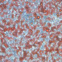
- 细胞名称:H22细胞(小鼠肝癌细胞)
- 形态:淋巴母细胞样,悬浮生长
- 含量:>1x10⁶ 个/瓶
- 污染:支原体、细菌、酵母和真菌检测为阴性
- 规格:T25瓶或者1mL冻存管包装
二、细胞接收后的处理:
1、贴壁细胞
- 收到T25方瓶细胞后,请检查是否漏液,如果漏液,请拍照片发给我们(冻存管细胞收到后直接37℃水浴复苏或直接放置于液氮中长期储存)。
- 请先在显微镜下确认细胞生长状态,去掉封口膜并将T25瓶置于37℃培养约2-3h。
- 弃去T25瓶中的培养基,换用新鲜的完全培养基。
- 如果细胞长满(90%以上)请及时进行细胞传代。
- 接到细胞次日,请检查细胞是否污染,若发现污染或疑似污染,请及时与我们取得联系。
2、悬浮细胞
- 收到细胞后,请检查是否漏液,如果漏液,请拍照片发给我们。
- 请先在显微镜下确认细胞生长状态,去掉封口膜并将15ml离心管置于37℃培养约2-3h。
- 1200rpm离心5min,弃去15ml离心管中的培养基,细胞沉淀用新鲜的完全培养基重悬并培养。
- 如果细胞长满(90%以上)请及时进行细胞传代。
- 接到细胞次日,请检查细胞是否污染,若发现污染或疑似污染,请及时与我们取得联系。
本公司的细胞培养操作规程,供参考
一、培养基及培养冻存条件准备:
- 准备RPMI-1640培养基,90%;优质胎牛血清,10%。
- 培养条件: 气相:空气,95%;二氧化碳,5%。 温度:37℃,培养箱湿度为70%-80%。
- 冻存液:90%血清,10%DMSO,现用现配。液氮储存。
对于贴壁细胞,传代可参考以下方法:
- 弃去培养上清,用不含钙、镁离子的PBS润洗细胞1-2次。
- 加2ml消化液(0.25%Trypsin-0.53mM EDTA)于培养瓶中,置于37℃培养箱中消化2-3分钟,然后在显微镜下观察细胞消化情况,若细胞大部分变圆并脱落,迅速拿回操作台,轻敲几下培养瓶后加入3ml此细胞的培养基终止消化。
- 轻轻吹打后吸出,移入15ml离心管中,在1200RPM条件下离心5分钟,弃去上清液,加入1mL培养液后吹匀。
- 移入到事先准备好的含有5ml培养基的T-25培养瓶中或含有14ml培养基的T-75培养瓶中培养。
3)细胞冻存:待细胞生长状态良好时,可进行细胞冻存。贴壁细胞冻存时,先要消化处理并进行细胞计数。消化方法按照细胞传代方法的1-3步骤进行,最后的重悬液使用血清。悬浮细胞直接计数后离心,用血清重悬浮,加DMSO至最终浓度为10%。加入DMSO后迅速混匀,按每1ml的数量分配到冻存管中。本公司按每个冻存管细胞数目大于1X10⁶个细胞冻存。
注意事项:
1. 收到冻存管细胞后,若发现干冰已挥发干净、冻存管瓶盖脱落、破损及细胞有污染,请立即与我们联系。
2. 所有动物细胞均视为有潜在的生物危害性,必须在二级生物安全台内操作,并请注意防护,所有废液及接触过此细胞的器皿需要灭菌后方能丢弃。
3. 细胞用途:仅供科研使用。
发货方式:
复苏后发货:我们复苏细胞后发货,货期一周左右,免运费。(气温较好建议复苏后发货)
冻存发货(干冰运输):需额外增加干冰运费,选择干冰运输的我们发两管细胞,为了保证客户接种可靠性多发一管。(气温低于0℃须冻存发货)
细胞发货采取专业的运输包装,并选择最快捷的运输方式(顺丰速运或其他空运快递)
Convolvulus Scammonia crude Alkaloids extract induces apoptosis through microtubules destruction in mice hepatoma H22 cell line
T his study evaluated the ability of crude alkaloids extracted from the leaves of Convolvulus Scammonia to distract the microtubule network of mice hepatocarcinoma cell line (H22), which is an invasive metastasis cell line. This assessment was carried out using the immunostaining technique. The extract was able to distract the microtubules of the cells under investigation after 60 min of exposure in a concentration as little as 20 g/ml. when DAPI staining used, the cells apoptosis was not detected in this concentration and time. The apoptotic cell have been observed when the concentration of the alkaloid extract elevated up to 80 and 100 g/ml during the mentioned exposure time. The cells were capable of recovering there na-tive microtubules constriction after 12 hr of the alkaloid removal from the media. The extract concentration of 1mg/Kg/Bw efficiently inhibited H22 cell line tumor growth in vivo to 97.14% in mice after three weeks treatment compared to untreated control animals. I n eukaryotic cells the cytoskeleton network consists of three major structural elements, microtubules, microfila-ment, and intermediate filaments (1). This network plays specific role in cell division, intracellular contacts, interac-tion with membranes, extracellular matrix, cell motion and maintenance or changes of cell shape (2). The diameter of microtubules (MTs) is about 25 nm they are composed of 13 equally spaced pro-filaments (2). Tubulin is the basic protein of the MTs, molecules of tubulin arranged in dimmers con-sisting of two forms, α-tubulin and β-tubulin. They are con-tinuously changeable structures (3), polymerization and de-polymerization of MTs is regulated by extra and intra-cellular factors (4). The presence of GTP at MTs ends is necessary to maintain the stability of the polymer (5). Because of their key role in cell function, microtubules emerged as important targets for cancer therapy. Taxanes and vinca alkaloids are microtubule inhibitors that destabilize microtubules, thereby suppressing their dynamics which required for proper mitotic function and effectively blocking cell cycle progression re-sulting in apoptosis. In spite of their antitumor activity, drug resistance to such MTs inhibitors is common, limiting their overall clinical efficacy. Therefore the discovery of novel agents that may overcome resistance to conventional MTs in-hibitors and provide higher efficacy of microtubule-targeting with limited toxicity is actually need (6). In addition, despite the success of taxanes and vinca alkaloids to inhibit the pro-gression of some cancers in clinical use, resistance to anti-microtubule agents is encountered in many tumor types, par-ticularly during multiple cycles of therapy. Therefore, there has been great interest in identifying and developing novel anti-microtubule drugs. (7) Moreover the most widely used Vinca alkaloids such as vinblastine, vincristine, and vindesine, often induce some in-tractable side effects including neurological and hematologi-cal toxicities and in particular, experience with both acquired Introduction.
Effect of Haimiding on the Functioning of Red Cell Membrane of FC and H22 Tumor-bearing Mice
风险提示:丁香通仅作为第三方平台,为商家信息发布提供平台空间。用户咨询产品时请注意保护个人信息及财产安全,合理判断,谨慎选购商品,商家和用户对交易行为负责。对于医疗器械类产品,请先查证核实企业经营资质和医疗器械产品注册证情况。
- 作者
- 内容
- 询问日期
 文献和实验
文献和实验Chicken intestinal epithelial cells were obtained from NEWGAINBIO company. Cells were cultured on 37℃, with 5% CO2, in the Ham’s F-12 Nutrient (DMEM/12) that contained the following supplementations: fetal bovine serum (5%), in-sulin (5 µg/mL), transferrin (5 µg/mL), selenium (5 ng/mL), epidermal growth factor (5 ng/mL) and penicillin-streptomycin (100–100 U/mL) for cell culturing (full DMEM/12). Experiments were performed with chicken intestinal epithelial cells and working solutions were prepared with plain DMEM/12 without supplementation. For the investigations, cells were seeded onto 96-well, 24-well or 6-well polystyrene cell culture plates.
Primary hVICs (passage 2) were cultured to 50–60% confluence and infected with pGMLV-SV40T-puro lentivirus (NewgainBio, Wuxi, China) at a multiplicity of infection of 80 supplemented with 5 µg/mL polybrene (Sigma-Aldrich, Buchs, Switzerland).
Tissue was cultured until cells became visible around the tissue, and when the fusion reached 90% (FIGURE 1A) §ask ¦lled with the prepared culturing medium was sent to the company for further immortalisation. Cell immortalisation was done for cell stability and longer-term use. Immortalised cells were cultured with 10% FBS and 1% PS in the DMEM medium. After the cells multiplied and merged, they were routinely passed and grown ( NEWGAINBIO Inc. Wuxi, Jiangsu, China) (FIGURE 1B-C).
Mouse primary cultured renal vascular ECs and VSMCs were obtained from Newgainbio company, which were tested by Factor VIII and α-smooth muscle actin (α-SMA), the marker of ECs and VSMCs. RNeasy Mini Kit was used for RNA extraction, and the above protocols were repeated.
Porcine primary colon epithelial cells (Newgainbio company, Wuxi,China) were cultured in Dulbecco's Modified Eagle's Medium (Solarbio, Beijing, China) containing 10 % fetal bovine serum (BioInd, Kiryat shmona, Lsrael) at 37 ◦C and 5 % CO2 humidity.
holybible1263 请问体外做hepG-2,体内做H22,有可比性吗? fuxiao29 我个人认为体外做hepG-2实验,体内也应该用同一种细胞系做,这样才有可比性。 holybible1263 谢谢,不过HEPG-2没有办法做体内呀,体内要用什么呢? fuxiao29 可以做体内实验呀,你可以将你用的质粒转染到HEPG-2细胞系里,构建稳定细胞系
。 2. 接种密度:传代比例 1:2 ~ 1:4,70% - 80% 汇合度时传代。 3. 控制胰酶消化时间:避免过长损伤细胞。 4. 冻存复苏温度要求:冻存缓慢降温,复苏快速升温,减少损伤。 5. 复苏后换液原则:24 小时后首次换液,避免频繁操作。 H22 细胞 小鼠肝癌细胞,分离自小鼠腹水,为悬浮细胞,呈淋巴母细胞样[10]。 培养条件:RPMI-1640培养基,10% 胎牛血清,1% 青霉素链霉素双抗[11]。 注意事项:
Nature 子刊:超声波调控肿瘤内细菌的基因表达,助力肿瘤免疫治疗
提高脾脏 M1 型巨噬细胞、CD4+T 细胞、CD8+T 细胞与记忆 T 细胞的占比,表明超声调控细菌表达 IFN-γ 能有效激活小鼠的系统性免疫(图 4)。 图 4. 超声调控细菌表达 IFN-γ 抑制远端肿瘤的生长与转移 为探讨超声调控细菌表达 IFN-γ 能否用于深部肿瘤的治疗,研究团队建立了小鼠原位移植肝肿瘤模型。在小鼠肝脏内接种 H22-Luc 细胞 4 天后,静脉注射超声响应性细菌,两天后采用超声辐照肿瘤,借助小动物活体成像观察原位肝肿瘤的生长情况,结果显示,超声调控细菌表达 IFN


