

- ¥2980
- LMAI Bio
- LM-0367G-FITC
- 中国/美国/欧洲
- 2025年07月10日
- IF=1:50-200
- Human
- Human

企业认证
相关产品推荐更多 >


万千商家帮你免费找货
0 人在求购买到急需产品
- 详细信息
- 文献和实验
- 技术资料
- 供应商:
上海联迈生物工程有限公司
- 库存:
大量
- 靶点:
详见说明书
- 级别:
1
- 目录编号:
LM-0367G-FITC
- 克隆性:
多克隆
- 抗原来源:
Rabbit
- 保质期:
1年
- 抗体英文名:
Anti-Complement C3/FITC
- 抗体名:
Anti-Complement C3/FITC
- 标记物:
FITC标记
- 宿主:
Human
- 适应物种:
Human
- 免疫原:
详见说明书
- 亚型:
IGg
- 形态:
粉末、液体、冻干粉
- 应用范围:
IF=1:50-200
- 浓度:
1mg/ml
- 保存条件:
-20 °C
- 规格:
100ul
| 英文名称 | Anti-Complement C3/FITC |
| 中文名称 | FITC标记的补体C3抗体 |
| 别 名 | Acylation stimulating protein cleavage product; Acylation-stimulating protein cleavage product; AHUS5; ARMD9; ASP; ASP; C3 and PZP-like alpha-2-macroglobulin domain-containing protein 1 antibody C3 antibody C3a anaphylatoxin; CO3_HUMAN; Complement C3 alpha chain; Complement C3; Complement C3 beta chain; Complement C3 precursor; Complement C3b alpha chain; Complement C3c alpha' chain fragment 2; Complement C3c fragment; Complement C3d fragment; Complement C3f fragment; Complement C3g fragment; Complement component 3; Complement component 3 precursor; Complement component C3; Complement factor 3; CPAMD1; Plp. |
| 规格价格 | 100ul/2980元 购买 大包装/询价 |
| 说 明 书 | 100ul |
| 研究领域 | 细胞生物 免疫学 神经生物学 细胞类型标志物 |
| 抗体来源 | Goat |
| 克隆类型 | Polyclonal |
| 交叉反应 | Human, |
| 产品应用 | IF=1:50-200 not yet tested in other applications. optimal dilutions/concentrations should be determined by the end user. |
| 分 子 量 | 187kDa |
| 性 状 | Lyophilized or Liquid |
| 浓 度 | 1mg/ml |
| 免 疫 原 | human Complement C3 purified from human plasma |
| 亚 型 | IgG |
| 纯化方法 | affinity purified by Protein A |
| 储 存 液 | 0.01M TBS(pH7.4) with 1% BSA, 0.03% Proclin300 and 50% Glycerol. |
| 保存条件 | Store at -20 °C for one year. Avoid repeated freeze/thaw cycles. The lyophilized antibody is stable at room temperature for at least one month and for greater than a year when kept at -20°C. When reconstituted in sterile pH 7.4 0.01M PBS or diluent of antibody the antibody is stable for at least two weeks at 2-4 °C. |
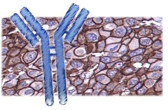
| 产品介绍 | background: The complement factor C3 consists of an alpha and a beta chain. C3 is a central factor in the complement cascade. It is central to the alternative pathway that leads to the C3 convertase C3bBb. The classical mannose binding lectin activation pathway leads to the C3 convertase C4b2a. These convertases cleave C3 resulting in C3a and C3b. Further degradation leads to the formation of the alpha chain products C3d, C3g and C3c. C3 is an acute phase protein that is produced by a wide range of tissues, including renal epithelial cells and hepatocytes. Function: C3 plays a central role in the activation of the complement system. Its processing by C3 convertase is the central reaction in both classical and alternative complement pathways. After activation C3b can bind covalently, via its reactive thioester, to cell surface carbohydrates or immune aggregates. Derived from proteolytic degradation of complement C3, C3a anaphylatoxin is a mediator of local inflammatory process. It induces the contraction of smooth muscle, increases vascular permeability and causes histamine release from mast cells and basophilic leukocytes. Acylation stimulating protein (ASP): adipogenic hormone that stimulates triglyceride (TG) synthesis and glucose transport in adipocytes, regulating fat storage and playing a role in postprandial TG clearance. Appears to stimulate TG synthesis via activation of the PLC, MAPK and AKT signaling pathways. Ligand for GPR77. Promotes the phosphorylation, ARRB2-mediated internalization and recycling of GPR77. Subunit: C3 precursor is first processed by the removal of 4 Arg residues, forming two chains, beta and alpha, linked by a disulfide bond. C3 convertase activates C3 by cleaving the alpha chain, releasing C3a anaphylatoxin and generating C3b (beta chain + alpha' chain). C3dg interacts with CR2 (via the N-terminal Sushi domains 1 and 2). During pregnancy, C3dg exists as a complex (probably a 2:2:2 heterohexamer) with AGT and the proform of PRG2. Interacts with VSIG4. C3b interacts with herpes simplex virus 1 (HHV-1) and herpes simplex virus 2 (HHV-2) envelope glycoprotein C; this interaction inhibits the activation of the complement system. Interacts with S.aureus immunoglobulin-binding protein sbi, this prevents interaction between C3dg and CR2. Interacts with S.aureus fib. Interacts (both C3a and ASP) with GPR77; the interaction occurs with higher affinity for ASP, enhancing the phosphorylation and activation of GPR77, recruitment of ARRB2 to the cell surface and endocytosis of GRP77. Subcellular Location: Secreted. Tissue Specificity: Plasma. The acylation stimulating protein (ASP) is expressed in adiopocytes and released into the plasma during both the fasting and postprandial periods. Post-translational modifications: C3b is rapidly split in two positions by factor I and a cofactor to form iC3b (inactivated C3b) and C3f which is released. Then iC3b is slowly cleaved (possibly by factor I) to form C3c (beta chain + alpha' chain fragment 1 + alpha' chain fragment 2), C3dg and C3f. Other proteases produce other fragments such as C3d or C3g. C3a is further processed by carboxypeptidases to release the C-terminal arginine residue generating the acylation stimulating protein (ASP). Levels of ASP are increased in adipocytes in the postprandial period and by insulin and dietary chylomicrons. Phosphorylation sites are present in the extracellular medium. DISEASE: Defects in C3 are the cause of complement component 3 deficiency (C3D) [MIM:613779]. A rare defect of the complement classical pathway. Patients develop recurrent, severe, pyogenic infections because of ineffective opsonization of pathogens. Some patients may also develop autoimmune disorders, such as arthralgia and vasculitic rashes, lupus-like syndrome and membranoproliferative glomerulonephritis. Genetic variation in C3 is associated with susceptibility to age-related macular degeneration type 9 (ARMD9) [MIM:611378]. ARMD is a multifactorial eye disease and the most common cause of irreversible vision loss in the developed world. In most patients, the disease is manifest as ophthalmoscopically visible yellowish accumulations of protein and lipid that lie beneath the retinal pigment epithelium and within an elastin-containing structure known as Bruch membrane. Defects in C3 are a cause of susceptibility to hemolytic uremic syndrome atypical type 5 (AHUS5) [MIM:612925]. An atypical form of hemolytic uremic syndrome. It is a complex genetic disease characterized by microangiopathic hemolytic anemia, thrombocytopenia, renal failure and absence of episodes of enterocolitis and diarrhea. In contrast to typical hemolytic uremic syndrome, atypical forms have a poorer prognosis, with higher death rates and frequent progression to end-stage renal disease. Note=Susceptibility to the development of atypical hemolytic uremic syndrome can be conferred by mutations in various components of or regulatory factors in the complement cascade system. Other genes may play a role in modifying the phenotype. Note=Increased levels of C3 and its cleavage product ASP, are associated with obesity, diabetes and coronary heart disease. Short-term endurance training reduces baseline ASP levels and subsequently fat storage. Similarity: Contains 1 anaphylatoxin-like domain. Contains 1 NTR domain. Database links: Entrez Gene: 718 Human Omim: 120700 Human SwissProt: P01024 Human Important Note: This product as supplied is intended for research use only, not for use in human, therapeutic or diagnostic applications. 补体(complement,C)是由近20种不同血清蛋白组成的多分子系统,约占血清球蛋白总量的10%。补体在血清中的含量相对稳定,不因免疫应答而增加,仅在某些病理情况下才会发生波动。补体系统的基本组成包括9种血清蛋白成分,按发现的先后顺序而分别命名为C1-9。C3是一种β球蛋白,是补体中含量最多、作用最重要的一个组分,是补体两条主要激活途径的中心环节,有重要的生物活性作用,可在肝脏中产生。C3蛋白由α、β2条肽链经二硫键连接而成,本产品能特异性的识别C3的β链。 |
风险提示:丁香通仅作为第三方平台,为商家信息发布提供平台空间。用户咨询产品时请注意保护个人信息及财产安全,合理判断,谨慎选购商品,商家和用户对交易行为负责。对于医疗器械类产品,请先查证核实企业经营资质和医疗器械产品注册证情况。
 文献和实验
文献和实验当FITC在碱性溶液中与抗体蛋白反应时,主要是蛋白质上赖氨酸的r氨基与荧光素的硫碳胺键(thiocarbmide)结合,形成FITC-蛋白质结合物,即荧光抗体或荧光结合物。一个IgG分子中有86个赖氨酸残基,一般最多能结合15~20个,一个IgG分子可结合2~8个分子的FITC,其反应式如下FITC-N=C=S + N-H2-蛋白质 → FITC-NS-C-N-H2-蛋白质常用Marsshall(1958)法标记荧光抗体,也可以根据条件采用Chadwick等标记法或Clark
ml三蒸水中即成; 方法与步骤: 根据Marshall氏法高效价的抗人球蛋白兔免疫血清,分离球蛋白。 1. 用0.15 mol/L NaCl的盐水及0.15 mol/L pH9.0的NaHCO3-Na2CO3缓冲液稀释使每毫升内含抗体10mg,缓冲液为总量的10%; 2. 将以上溶液降温至4℃,按蛋白:荧光素=50—80mg:1mg的比例加入异硫氰酸荧光素,在0—4℃下电磁搅拌12—14h; 3.用半饱和硫酸铵将标记球蛋白
5. 过柱。取透析过夜的标记物,过葡萄糖凝胶G-25或G-50柱,分离出游离荧光素,收集标记的荧光抗体进行鉴定。
 技术资料
技术资料暂无技术资料 索取技术资料

