


- ¥2980
- LMAI Bio
- LM-5185R-FITC
- 中国/美国/欧洲
- 2025年07月09日
- ICC=1:50-200 IF=1:50-200
- Human, Mouse, Rat, Chicken, Dog, Pig, Cow, Sheep,
- Human, Mouse, Rat, Chicken, Dog, Pig, Cow, Sheep,

企业认证
相关产品推荐更多 >


万千商家帮你免费找货
0 人在求购买到急需产品
- 详细信息
- 文献和实验
- 技术资料
- 供应商:
上海联迈生物工程有限公司
- 库存:
大量
- 靶点:
详见说明书
- 级别:
1
- 目录编号:
LM-5185R-FITC
- 克隆性:
多克隆
- 抗原来源:
Rabbit
- 保质期:
1年
- 抗体英文名:
Anti-phospho-AQP2 (Ser264)/FITC
- 抗体名:
Anti-phospho-AQP2 (Ser264)/FITC
- 标记物:
FITC标记
- 宿主:
Human, Mouse, Rat, Chicken, Dog, Pig, Cow, Sheep,
- 适应物种:
Human, Mouse, Rat, Chicken, Dog, Pig, Cow, Sheep,
- 免疫原:
详见说明书
- 亚型:
IGg
- 形态:
粉末、液体、冻干粉
- 应用范围:
ICC=1:50-200 IF=1:50-200
- 浓度:
1mg/ml
- 保存条件:
-20 °C
- 规格:
100ul
| 英文名称 | Anti-phospho-AQP2 (Ser264)/FITC |
| 中文名称 | FITC标记的磷酸化水通道蛋白2抗体 |
| 别 名 | ADH water channel; AQP 2; AQP CD; AQP2; AQPCD; Aquaporin 2 collecting duct; Aquaporin CD; Aquaporin2; Aquaporine 2; Collecting duct water channel protein; MGC34501; Water channel protein for renal collecting duct; WCH CD; WCHCD. |
| 规格价格 | 100ul/2980元 购买 大包装/询价 |
| 说 明 书 | 100ul |
| 产品类型 | 磷酸化抗体 |
| 研究领域 | 肿瘤 免疫学 信号转导 通道蛋白 细胞粘附分子 |
| 抗体来源 | Rabbit |
| 克隆类型 | Polyclonal |
| 交叉反应 | Human, Mouse, Rat, Chicken, Dog, Pig, Cow, Sheep, |
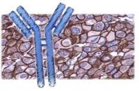
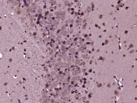
| 产品应用 | ICC=1:50-200 IF=1:50-200 not yet tested in other applications. optimal dilutions/concentrations should be determined by the end user. |
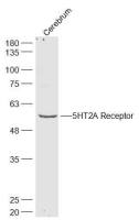
| 分 子 量 | 30kDa |
| 细胞定位 | 细胞膜 |
| 性 状 | Lyophilized or Liquid |
| 浓 度 | 1mg/ml |
| 免 疫 原 | KLH conjugated Synthesised phosphopeptide derived from human AQP2 around the phosphorylation site of Ser264 |
| 亚 型 | IgG |
| 纯化方法 | affinity purified by Protein A |
| 储 存 液 | 0.01M TBS(pH7.4) with 1% BSA, 0.03% Proclin300 and 50% Glycerol. |
| 保存条件 | Store at -20 °C for one year. Avoid repeated freeze/thaw cycles. The lyophilized antibody is stable at room temperature for at least one month and for greater than a year when kept at -20°C. When reconstituted in sterile pH 7.4 0.01M PBS or diluent of antibody the antibody is stable for at least two weeks at 2-4 °C. |
| 产品介绍 | background: This gene encodes a water channel protein located in the kidney collecting tubule. It belongs to the MIP/aquaporin family, some members of which are clustered together on chromosome 12q13. Mutations in this gene have been linked to autosomal dominant, and recessive forms of nephrogenic diabetes insipidus. Belongs to the MIP/aquaporin (TC 1.A.8) family. Function: Forms a water-specific channel that provides the plasma membranes of renal collecting duct with high permeability to water, thereby permitting water to move in the direction of an osmotic gradient. Subcellular Location: Apical cell membrane; Multi-pass membrane protein. Cytoplasmic vesicle membrane; Multi-pass membrane protein. Note=Shuttles from vesicles to the apical membrane. Tissue Specificity: Expressed in renal collecting tubules. Post-translational modifications: Ser-256 phosphorylation is necessary and sufficient for expression at the apical membrane. Endocytosis is not phosphorylation-dependent. DISEASE: Defects in AQP2 are the cause of diabetes insipidus nephrogenic autosomal (ANDI) [MIM:125800]; also known as diabetes insipidus nephrogenic type 2. ANDI is caused by the inability of the renal collecting ducts to absorb water in response to arginine vasopressin. It is characterized by excessive water drinking (polydypsia), excessive urine excretion (polyuria), persistent hypotonic urine, and hypokalemia. Inheritance can be autosomal dominant or recessive. Similarity: Belongs to the MIP/aquaporin (TC 1.A.8) family. Database links: Entrez Gene: 359 Human Entrez Gene: 11827 Mouse Entrez Gene: 25386 Rat Omim: 107777 Human SwissProt: P41181 Human SwissProt: P56402 Mouse SwissProt: P34080 Rat Unigene: 130730 Human Unigene: 20206 Mouse Unigene: 90076 Rat Important Note: This product as supplied is intended for research use only, not for use in human, therapeutic or diagnostic applications. |
风险提示:丁香通仅作为第三方平台,为商家信息发布提供平台空间。用户咨询产品时请注意保护个人信息及财产安全,合理判断,谨慎选购商品,商家和用户对交易行为负责。对于医疗器械类产品,请先查证核实企业经营资质和医疗器械产品注册证情况。
 文献和实验
文献和实验,如异硫氰酸荧光素(fluorescein isothiocyanate,FITC)将细胞膜蛋白全部进行标记。也可用特异性的探针,如荧光抗体,标记特异的膜蛋白。膜蛋白一旦被标记就可用激光束进行局部照射处理,使荧光脱色,形成直径约为1μm的白斑。若是可移动的膜蛋白,则会因蛋白的移动,使白斑消失,若是不能移动的蛋白.则白斑不会消失。根据荧光恢复的速度, 可推算膜脂的扩散速度为每秒钟为几个微米,而膜蛋白的扩散速度变化幅度较大,少数膜蛋白的扩散速度可达到膜脂的速度,大多数蛋白的扩散速度都比膜脂慢,还有一些膜蛋白
荧光素(FITC)标记的TFAR19单克隆抗体为探针,对细胞凋亡过程中TFAR19蛋白的表达水平及定位研究发现,凋亡早期TFAR19表达水平增高并出现快速核转位现象,伴随着细胞核形态学的变化,持续较长时间,在凋亡小体中仍然可见。同时我们发现,凋亡早期TFAR19蛋白的核转位早于磷脂酰丝氨酸(PS)外翻和细胞核DNA的片段化,提示TFAR19蛋白的核转位是细胞凋亡更早期发生的事件之一。进一步的研究证明,凋亡早期TFAR19的核转位具有普遍意义,不同细胞凋亡早期均出现TFAR19高表达和核转
生长、分化、成熟变化、细胞的三维结构、染色体分析、基因表达、基因诊断。changqing112:我在实验中设想利用激光共聚焦显微镜来观测单个细胞中的通道蛋白分布情况,不知需要的抗体有何要求?必须是单克隆抗体吗?如果抗体是普通的抗体,那该如何检测呢? 土人:你说的是免疫荧光染色的方法吧? 免疫荧光染色跟免疫组化差不多,差别就在于前者是用的荧光染料(如cy2、cy3、FITC等)标记的二抗,在荧光显微镜或者激光共聚焦显微镜下观察荧光;而后者则是用的直接显色的方法,在普通光镜下或者肉眼下直接就可以看到信号
 技术资料
技术资料暂无技术资料 索取技术资料

