

- ¥780 - 2200
- LMAI Bio
- LM-6741R
- 进口/国产
- 2026年02月09日
- WB=1:500-2000 ELISA=1:500-1000 IHC-P=1:400-800 IHC-F=1:400-800 Flow-Cyt=1ug/test IF=1:100-500 (石蜡切片需做抗原修复)
- Rabbit
- Human, Mouse, Rat, Dog, Pig, Cow, Horse, Rabbit,

企业认证
相关产品推荐更多 >


万千商家帮你免费找货
0 人在求购买到急需产品
- 详细信息
- 文献和实验
- 技术资料
- 供应商:
上海联迈生物工程有限公司
- 库存:
大量
- 目录编号:
LM-6741R
- 克隆性:
多克隆
- 抗原来源:
Rabbit
- 保质期:
1年
- 抗体英文名:
ASC
- 抗体名:
凋亡相关斑点样蛋白ASC抗体
- 宿主:
Rabbit
- 适应物种:
Human, Mouse, Rat, Dog, Pig, Cow, Horse, Rabbit,
- 免疫原:
KLH conjugated synthetic peptide derived from human TMS1/ASC:31-130/195
- 亚型:
IgG
- 形态:
Lyophilized or Liquid
- 应用范围:
WB=1:500-2000 ELISA=1:500-1000 IHC-P=1:400-800 IHC-F=1:400-800 Flow-Cyt=1ug/test IF=1:100-500 (石蜡切片需做抗原修复)
- 浓度:
1mg/ml
- 保存条件:
Store at -20 °C
- 规格:
50ul 100ul 200ul
| 英文名称 | ASC |
| 中文名称 | 凋亡相关斑点样蛋白ASC抗体 |
| 别 名 | TMS1; Apoptosis associated speck like protein containing a CARD; Apoptosis-associated speck-like protein containing a CARD; ASC_HUMAN; CARD 5; CARD5; Caspase recruitment domain containing protein 5; Caspase recruitment domain protein 5; Caspase recruitment domain-containing protein 5; hASC; MGC10332; PYCARD; TMS-1; PYD and CARD domain containing; PYD and CARD domain containing protein; PYD and CARD domain-containing protein; Target of methylation induced silencing 1; Target of methylation-induced silencing 1; TMS 1. |
 |
Specific References (1) | bs-6741R has been referenced in 1 publications. [IF=3.06] Zhang, Bo, et al. "Cortistatin inhibits NLRP3 inflammasome activation of cardiac fibroblasts during sepsis." Journal of Cardiac Failure (2015). WB ; Rat. PubMed:25639691 |
| 规格价格 | 50ul/780元 购买 100ul/1380元 购买 200ul/2200元 购买 大包装/询价 |
| 说 明 书 | 50ul 100ul 200ul |
| 研究领域 | 肿瘤 细胞生物 免疫学 信号转导 细胞凋亡 转录调节因子 肿瘤细胞生物标志物 |
| 抗体来源 | Rabbit |
| 克隆类型 | Polyclonal |
| 交叉反应 | Human, Mouse, Rat, Dog, Pig, Cow, Horse, Rabbit, |
| 产品应用 | WB=1:500-2000 ELISA=1:500-1000 IHC-P=1:400-800 IHC-F=1:400-800 Flow-Cyt=1ug/test IF=1:100-500 (石蜡切片需做抗原修复) not yet tested in other applications. optimal dilutions/concentrations should be determined by the end user. |
| 分 子 量 | 22kDa |
| 细胞定位 | 细胞核 细胞浆 |
| 性 状 | Lyophilized or Liquid |
| 浓 度 | 1mg/ml |
| 免 疫 原 | KLH conjugated synthetic peptide derived from human TMS1/ASC:31-130/195 |
| 亚 型 | IgG |
| 纯化方法 | affinity purified by Protein A |
| 储 存 液 | 0.01M TBS(pH7.4) with 1% BSA, 0.03% Proclin300 and 50% Glycerol. |
| 保存条件 | Store at -20 °C for one year. Avoid repeated freeze/thaw cycles. The lyophilized antibody is stable at room temperature for at least one month and for greater than a year when kept at -20°C. When reconstituted in sterile pH 7.4 0.01M PBS or diluent of antibody the antibody is stable for at least two weeks at 2-4 °C. |
| PubMed | PubMed |
| 产品介绍 | background: Promotes caspase-mediated apoptosis. This proapoptotic activity is mediated predominantly through the activation of caspase-9. May be a component of the inflammasome, a protein complex which also includes NALP2, CARD8 and CASP1 and whose function would be the activation of proinflammatory caspases. Tissue specificity: Widely expressed at low levels. Detected in peripheral blood leukocytes, lung, small intestine, spleen, thymus, colon and at lower levels in placenta, liver and kidney. Very low expression in skeletal muscle, heart and brain. Detected in the leukemia cell lines HL-60 and U937, but not in Jurkat T-cell lymphoma and Daudi Burkitt's lymphoma. Function: Promotes caspase-mediated apoptosis. This proapoptotic activity is mediated predominantly through the activation of caspase-9. May be a component of the inflammasome, a protein complex which also includes NALP2, CARD8 and CASP1 and whose function would be the activation of proinflammatory caspases. Subunit: Forms complexes with other DAPIN domain-containing proteins. Interacts with CIAS1/PYPAF1 and PYDC1. Subcellular Location: Cytoplasm. Note=Upstream of caspase activation, a redistribution from the cytoplasm to the aggregates occurs. These appear as hollow, perinuclear spherical, ball-like structures. Tissue Specificity: Widely expressed at low levels. Detected in peripheral blood leukocytes, lung, small intestine, spleen, thymus, colon and at lower levels in placenta, liver and kidney. Very low expression in skeletal muscle, heart and brain. Detected in the leukemia cell lines HL-60 and U937, but not in Jurkat T-cell lymphoma and Daudi Burkitt's lymphoma. Detected in the melanoma cell line WM35, but not in WM793. Not detected in HeLa cervical carcinoma cells and Molt 4 lymphocytic leukemia cells. Post-translational modifications: Phosphorylated. Similarity: Contains 1 CARD domain. Contains 1 DAPIN domain. SWISS: Q9ULZ3 Gene ID: 29108 Database links: Entrez Gene: 29108 Human Omim: 606838 Human SwissProt: Q9ULZ3 Human Unigene: 499094 Human Important Note: This product as supplied is intended for research use only, not for use in human, therapeutic or diagnostic applications. |
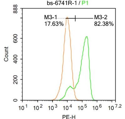
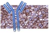
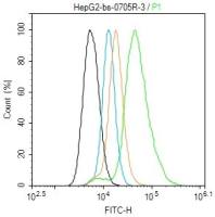
| 产品图片 |  Sample:Spinal cord (Rat)Lysate at 40 ug Primary: Anti-ASC(bs-6741R)at 1/300 dilution Secondary: IRDye800CW Goat Anti-RabbitIgG at 1/20000 dilution Predicted band size: 22kD Observed band size: 18kD 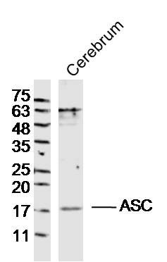 Sample:Cerebrum (Rat)Lysate at 40 ug Primary: Anti-ASC(bs-6741R)at 1/300 dilution Secondary: IRDye800CW Goat Anti-RabbitIgG at 1/20000 dilution Predicted band size: 22kD Observed band size: 18kD  Paraformaldehyde-fixed, paraffin embedded (human gastric carcinoma); Antigen retrieval by boiling in sodium citrate buffer (pH6.0) for 15min; Block endogenous peroxidase by 3% hydrogen peroxide for 20 minutes; Blocking buffer (normal goat serum) at 37°C for 30min; Antibody incubation with (ASC) Polyclonal Antibody, Unconjugated (bs-6741R) at 1:400 overnight at 4°C, followed by operating according to SP Kit(Rabbit) (sp-0023) instructionsand DAB staining.  Paraformaldehyde-fixed, paraffin embedded (mouse brain tissue); Antigen retrieval by boiling in sodium citrate buffer (pH6.0) for 15min; Block endogenous peroxidase by 3% hydrogen peroxide for 20 minutes; Blocking buffer (normal goat serum) at 37°C for 30min; Antibody incubation with (ASC) Polyclonal Antibody, Unconjugated (bs-6741R) at 1:400 overnight at 4°C, followed by operating according to SP Kit(Rabbit) (sp-0023) instructionsand DAB staining. 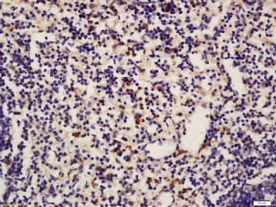 Tissue/cell: Mouse spleen tissue; 4% Paraformaldehyde-fixed and paraffin-embedded; Antigen retrieval: citrate buffer ( 0.01M, pH 6.0 ), Boiling bathing for 15min; Block endogenous peroxidase by 3% Hydrogen peroxide for 30min; Blocking buffer (normal goat serum,C-0005) at 37℃ for 20 min; Incubation: Anti-ASC Polyclonal Antibody, Unconjugated(bs-6741R) 1:200, overnight at 4°C, followed by conjugation to the secondary antibody(SP-0023) and DAB(C-0010) staining  Blank control:A549. Primary Antibody (green line): Rabbit Anti-ASC antibody (bs-6741R) Dilution: 1μg /10^6 cells; Isotype Control Antibody (orange line): Rabbit IgG . Secondary Antibody : Goat anti-rabbit IgG-PE Dilution: 1μg /test. Protocol The cells were fixed with 4% PFA (10min at room temperature)and then permeabilized with 20% PBST for 20 min at-20℃. The cells were then incubated in 5%BSA to block non-specific protein-protein interactions for 30 min at at room temperature .Cells stained with Primary Antibody for 30 min at room temperature. The secondary antibody used for 40 min at room temperature. Acquisition of 20,000 events was performed. |
风险提示:丁香通仅作为第三方平台,为商家信息发布提供平台空间。用户咨询产品时请注意保护个人信息及财产安全,合理判断,谨慎选购商品,商家和用户对交易行为负责。对于医疗器械类产品,请先查证核实企业经营资质和医疗器械产品注册证情况。
 文献和实验
文献和实验炎症促发阿尔茨海默病又添一力证!NLRP3 炎症小体激活引发 tau 蛋白异常
,Tau22 小鼠促炎因子相关基因(Jun,Fas 和 Toll 样受体)和染色质重构相关基因(Hdac2)存在相互作用,提示在 Tau 蛋白病早期,神经胶质细胞存在炎症表型和表观遗传层面的调控。同时,Tau22 小鼠胶质细胞存在明显形态学异常。图片来源:nature为进一步验证 NLRP3 炎症小体参与 tau 蛋白病的机制,研究人员分别构建了人细胞凋亡相关斑点样蛋白 Pycard 敲除(Tau22 /Asc−/−)和 Cias1 敲除(Tau22 /Nlrp3−/−)小鼠。实验结果表明 Tau22
通路,诱导多种 NOD 样受体(例如 NLRP3)蛋白以及 pro-IL-1β/pro-IL-18 的表达;次级信号为活化信号,细胞内的 NOD 样受体在迅速识别危险相关分子模式或病原体相关分子模式(例如线粒体 DNA)后,与接头蛋白 ASC 组装,从而招募 pro-Caspase-1[4]。当 pro-Caspase-1 的局部浓度升高时,发生自体剪切,生成的片段 p20 和 p10 组成四聚体结构的具有生物活性的 Caspase-1(即活化的 Caspase-1)[5]。炎症小体通过调控
「铲屎官」注意了!剑桥大学揭示这些基因的改变或让猫猫狗狗成为
导读2002 年,Tschopp 研究团队首次提出炎症小体(Inflammasome)的概念,此后炎症小体成为炎症疾病和免疫学领域的热门研究对象。十几年来,关于炎症小体的研究论文如雨后春笋层出不穷。先天免疫系统中,病原体入侵免疫细胞,进而启动机体炎症防御反应,最终发挥抗感染作用。炎症小体作为抵抗感染和肿瘤发生的关键机器,是一种相对较大的多聚体蛋白复合物,主要由传感器 NLRs(Nod-like receptors)、衔接蛋白 ASC(Apoptosis-associated speck
 技术资料
技术资料暂无技术资料 索取技术资料

