


- ¥1380 - 2200
- LMAI Bio
- LM-5718R
- 进口/国产
- 2026年01月07日
- WB=1:500-2000 ELISA=1:500-1000 IHC-P=1:400-800 IHC-F=1:400-800 IF=1:100-500 (石蜡切片需做抗原修复)
- Rabbit
- Human, Mouse, Rat, Dog, Pig, Cow, Rabbit, Sheep,

企业认证
相关产品推荐更多 >


万千商家帮你免费找货
0 人在求购买到急需产品
- 详细信息
- 文献和实验
- 技术资料
- 供应商:
上海联迈生物工程有限公司
- 库存:
大量
- 目录编号:
LM-5718R
- 克隆性:
多克隆
- 抗原来源:
Rabbit
- 保质期:
1年
- 抗体英文名:
Ensconsin
- 抗体名:
上皮细胞微管相关蛋白7抗体
- 宿主:
Rabbit
- 适应物种:
Human, Mouse, Rat, Dog, Pig, Cow, Rabbit, Sheep,
- 免疫原:
KLH conjugated synthetic peptide derived from human Ensconsin:151-250/749
- 亚型:
IgG
- 形态:
Lyophilized or Liquid
- 应用范围:
WB=1:500-2000 ELISA=1:500-1000 IHC-P=1:400-800 IHC-F=1:400-800 IF=1:100-500 (石蜡切片需做抗原修复)
- 浓度:
1mg/ml
- 保存条件:
Store at -20 °C
- 规格:
100ul 200ul
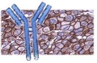
| 英文名称 | Ensconsin |
| 中文名称 | 上皮细胞微管相关蛋白7抗体 |
| 别 名 | E MAP 115; EMAP115; Epithelial microtubule associated protein of 115 kDa; MAP 7; MAP7; Microtubule associated protein 7; MAP7_HUMAN. |
| 规格价格 | 100ul/1380元 购买 200ul/2200元 购买 大包装/询价 |
| 说 明 书 | 100ul 200ul |
| 研究领域 | 肿瘤 细胞生物 免疫学 发育生物学 信号转导 干细胞 |
| 抗体来源 | Rabbit |
| 克隆类型 | Polyclonal |
| 交叉反应 | Human, Mouse, Rat, Dog, Pig, Cow, Rabbit, Sheep, |
| 产品应用 | WB=1:500-2000 ELISA=1:500-1000 IHC-P=1:400-800 IHC-F=1:400-800 IF=1:100-500 (石蜡切片需做抗原修复) not yet tested in other applications. optimal dilutions/concentrations should be determined by the end user. |
| 分 子 量 | 84kDa |
| 细胞定位 | 细胞核 细胞浆 细胞膜 |
| 性 状 | Lyophilized or Liquid |
| 浓 度 | 1mg/ml |
| 免 疫 原 | KLH conjugated synthetic peptide derived from human Ensconsin:151-250/749 |
| 亚 型 | IgG |
| 纯化方法 | affinity purified by Protein A |
| 储 存 液 | 0.01M TBS(pH7.4) with 1% BSA, 0.03% Proclin300 and 50% Glycerol. |
| 保存条件 | Store at -20 °C for one year. Avoid repeated freeze/thaw cycles. The lyophilized antibody is stable at room temperature for at least one month and for greater than a year when kept at -20°C. When reconstituted in sterile pH 7.4 0.01M PBS or diluent of antibody the antibody is stable for at least two weeks at 2-4 °C. |
| PubMed | PubMed |
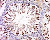
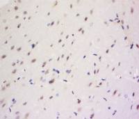
| 产品介绍 | background: Ensconsin is a microtubule associated protein that is predominantly expressed in cells of epithelial origin. Microtubule associated proteins are thought to be involved in microtubule dynamics, which is essential for cell polarization and differentiation. This protein has been shown to be able to stabilize microtubules, and may serve to modulate microtubule functions. Studies of the related mouse protein also suggested an essential role in microtubule function required for spermatogenesis. Function: Microtubule-stabilizing protein that may play an important role during reorganization of microtubules during polarization and differentiation of epithelial cells. Associates with microtubules in a dynamic manner. May play a role in the formation of intercellular contacts. Colocalization with TRPV4 results in the redistribution of TRPV4 toward the membrane and may link cytoskeletal microfilaments. Subunit: Interacts with TRPV4 (By similarity). Subcellular Location: Cytoplasm, perinuclear region. Basolateral cell membrane. Cytoplasm, cytoskeleton. Note=Colocalized on microtubules. An intracellular redistribution is triggered during induction of keratinocyte terminal differentiation from microtubules with a perinuclear localization to cortical microtubules organized in spike-like bundles facing intercellular contacts. Tissue Specificity: Expressed in the skin and cells of epithelial origin. Predominantly expressed in the suprabasal layers of the normal epidermis and relatively abundant in squamous cell carcinomas but barely detectable in basal cell carcinomas. Post-translational modifications: The association with microtubules is regulated by phosphorylation during the cell cycle. During interphase only phosphorylated on serine. Phosphorylated on threonine in mitosis. Similarity: Belongs to the MAP7 family. SWISS: Q14244 Gene ID: 9053 Database links: Entrez Gene: 9053 Human Entrez Gene: 17761 Mouse Omim: 604108 Human SwissProt: Q14244 Human SwissProt: O88735 Mouse Unigene: 486548 Human Unigene: 20928 Mouse Important Note: This product as supplied is intended for research use only, not for use in human, therapeutic or diagnostic applications. |
风险提示:丁香通仅作为第三方平台,为商家信息发布提供平台空间。用户咨询产品时请注意保护个人信息及财产安全,合理判断,谨慎选购商品,商家和用户对交易行为负责。对于医疗器械类产品,请先查证核实企业经营资质和医疗器械产品注册证情况。
 文献和实验
文献和实验免疫组织化学技术(immunohistochemistry)简介
7 确定肿瘤分期 判断肿瘤是原位还是浸润及有无血管、淋巴管侵袭与肿瘤分期密切相关。用常规病理方法判断有时是十分困难的,但用免疫组化法可获得明确结果。如采用层粘连蛋白和Ⅳ型胶原的单克隆抗体可清楚显示基底膜的主要成分,一旦证实上皮性癌突破了基底膜,就不是原位癌,而是浸润癌了,其预后是不同的。用第八因子相关蛋白、荆豆凝集素等显示血管和淋巴管内皮细胞的标记物则可清楚显示肿瘤对血管或淋巴管的浸润。对许多肿瘤的良恶性鉴别及有无血管或淋巴管浸润,这是主要的鉴别依据,同时也有治疗及预后意义。
胰腺上皮内瘤变和胰腺癌组织中TGF-f3/Smad信号通路相关蛋白表达
内瘤变(pancreaticintraepithelial neoplasia,PanlN)是胰腺癌的癌前病变,它反映了胰腺小导管上皮细胞从不典型增生至原位癌这一系列病变的连续过程 。有研究显示在此过程中也存在着TGF-13/Smads信号传导通路的异常 ],但因研究仅针对TGF-13/Smads信号传导通路中的个别元件,对于整个TGF-131/Smads信号传导通路的变化规律尚缺乏深入的认识。本研究用组织芯片和免疫组化技术系统地比较研究了不同级别PanIN和胰腺癌组织TGF-13/Smad信号传导通路中相关蛋白的表达,并联系临床病理资料作相关
性肌母细胞瘤,曾被认为是肌源性的,但该肿瘤肌源性标记阴性,而神经性标记阳性,证明为神经来源(可能来自神经鞘细胞)。分化很差的肿瘤病理上常按细胞形态分为梭形细胞肿瘤、小圆细胞肿瘤等,通过多种标记的联合应用,也可能确定来源。7、确定肿瘤分期判断肿瘤是原位还是浸润及有无血管、淋巴管侵袭与肿瘤分期密切相关。用常规病理方法判断有时是十分困难的,但用免疫组化法可获得明确结果。如采用层粘连蛋白和Ⅳ型胶原的单克隆抗体可清楚显示基底膜的主要成分,一旦证实上皮性癌突破了基底膜,就不是原位癌,而是浸润癌了,其预后是不同的。用第八因子相关蛋白
 技术资料
技术资料暂无技术资料 索取技术资料
