

- ¥1380 - 2200
- LMAI Bio
- LM-20487R
- 进口/国产
- 2026年03月19日
- WB=1:500-2000 ELISA=1:500-1000 IHC-P=1:400-800 IHC-F=1:400-800 ICC=1:100-500 IF=1:100-500 (石蜡切片需做抗原修复)
- Rabbit
- Human, Mouse,

企业认证
相关产品推荐更多 >


万千商家帮你免费找货
0 人在求购买到急需产品
- 详细信息
- 询价记录
- 文献和实验
- 技术资料
- 供应商:
上海联迈生物工程有限公司
- 库存:
大量
- 目录编号:
LM-20487R
- 克隆性:
多克隆
- 抗原来源:
Rabbit
- 保质期:
1年
- 抗体英文名:
PODXL
- 抗体名:
足细胞特异蛋白抗体
- 宿主:
Rabbit
- 适应物种:
Human, Mouse,
- 免疫原:
KLH conjugated synthetic peptide derived from human PODXL:401-500/558 <Extracellular>
- 亚型:
IgG
- 形态:
Lyophilized or Liquid
- 应用范围:
WB=1:500-2000 ELISA=1:500-1000 IHC-P=1:400-800 IHC-F=1:400-800 ICC=1:100-500 IF=1:100-500 (石蜡切片需做抗原修复)
- 浓度:
1mg/ml
- 保存条件:
Store at -20 °C
- 规格:
100ul 200ul
| 英文名称 | PODXL |
| 中文名称 | 足细胞特异蛋白抗体 |
| 别 名 | Podocalyxin; PCLP; PCLP1; podocalyxin-like isoform 1 precursor; Podocalyxin like protein; PODXL_HUMAN; GCTM-2 antigen; Gp200; Podocalyxin-like protein 1; PC; PCLP-1; PCX; PODXL. |
| 规格价格 | 100ul/1380元 购买 200ul/2200元 购买 大包装/询价 |
| 说 明 书 | 100ul 200ul |
| 研究领域 | 细胞生物 |
| 抗体来源 | Rabbit |
| 克隆类型 | Polyclonal |
| 交叉反应 | Human, Mouse, |
| 产品应用 | WB=1:500-2000 ELISA=1:500-1000 IHC-P=1:400-800 IHC-F=1:400-800 ICC=1:100-500 IF=1:100-500 (石蜡切片需做抗原修复) not yet tested in other applications. optimal dilutions/concentrations should be determined by the end user. |
| 分 子 量 | 59kDa |
| 细胞定位 | 细胞膜 |
| 性 状 | Lyophilized or Liquid |
| 浓 度 | 1mg/ml |
| 免 疫 原 | KLH conjugated synthetic peptide derived from human PODXL:401-500/558 <Extracellular> |
| 亚 型 | IgG |
| 纯化方法 | affinity purified by Protein A |
| 储 存 液 | 0.01M TBS(pH7.4) with 1% BSA, 0.03% Proclin300 and 50% Glycerol. |
| 保存条件 | Store at -20 °C for one year. Avoid repeated freeze/thaw cycles. The lyophilized antibody is stable at room temperature for at least one month and for greater than a year when kept at -20°C. When reconstituted in sterile pH 7.4 0.01M PBS or diluent of antibody the antibody is stable for at least two weeks at 2-4 °C. |
| PubMed | PubMed |
| 产品介绍 | background: This gene encodes a member of the sialomucin protein family. The encoded protein was originally identified as an important component of glomerular podocytes. Podocytes are highly differentiated epithelial cells with interdigitating foot processes covering the outer aspect of the glomerular basement membrane. Other biological activities of the encoded protein include: binding in a membrane protein complex with Na+/H+ exchanger regulatory factor to intracellular cytoskeletal elements, playing a role in hematopoetic cell differentiation, and being expressed in vascular endothelium cells and binding to L-selectin. [provided by RefSeq, Jul 2008] Function: Involved in the regulation of both adhesion and cell morphology and cancer progression. Function as an anti-adhesive molecule that maintains an open filtration pathway between neighboring foot processes in the podocyte by charge repulsion. Acts as a pro-adhesive molecule, enhancing the adherence of cells to immobilized ligands, increasing the rate of migration and cell-cell contacts in an integrin-dependent manner. Induces the formation of apical actin-dependent microvilli. Involved in the formation of a preapical plasma membrane subdomain to set up inital epithelial polarization and the apical lumen formation during renal tubulogenesis. Plays a role in cancer development and aggressiveness by inducing cell migration and invasion through its interaction with the actin-binding protein EZR. Affects EZR-dependent signaling events, leading to increased activities of the MAPK and PI3K pathways in cancer cells. Subcellular Location: Apical cell membrane. Cell projection, lamellipodium. Cell projection, filopodium. Cell projection, ruffle. Cell projection, microvillus. Membrane raft. Membrane. In single attached epithelial cells is restricted to a preapical pole on the free plasma membrane whereas other apical and basolateral proteins are not yet polarized. Colocalizes with SLC9A3R2 at the apical plasma membrane during epithelial polarization. Colocalizes with SLC9A3R1 at the trans-Golgi network (transiently) and at the apical plasma membrane. Its association with the membrane raft is transient. Colocalizes with actin filaments, EZR and SLC9A3R1 in a punctate pattern at the apical cell surface where microvilli form. Colocalizes with EZR and SLC9A3R2 at the apical cell membrane of glomerular epithelium cells (By similarity). Forms granular, punctuated pattern, forming patches, preferentially adopting a polar distribution, located on the migrating poles of the cell or forming clusters along the terminal ends of filipodia establishing contact with the endothelial cells. Colocalizes with the submembrane actin of lamellipodia, particularly associated with ruffles. Colocalizes with vinculin at protrusions of cells. Colocalizes with ITGB1. Colocalizes with PARD3, PRKCI, EXOC5, OCLN, RAB11A and RAB8A in apical membrane initiation sites (AMIS) during the generation of apical surface and luminogenesis. Tissue Specificity: Glomerular epithelium cell (podocyte). Similarity: Belongs to the podocalyxin family. SWISS: O00592 Gene ID: 5420 Database links: Entrez Gene: 5420 Human Entrez Gene: 27205 Mouse Entrez Gene: 482252 Dog Omim: 602632 Human SwissProt: Q52S86 Dog SwissProt: O00592 Human SwissProt: Q9R0M4 Mouse Unigene: 732423 Human Unigene: 89918 Mouse Important Note: This product as supplied is intended for research use only, not for use in human, therapeutic or diagnostic applications. |
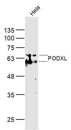
| 产品图片 |  Sample: Hela(Human) Cell Lysate at 40 ug Primary: Anti-PODXL (bs-20487R) at 1/300 dilution Secondary: IRDye800CW Goat Anti-Rabbit IgG at 1/20000 dilution Predicted band size: 59 kD Observed band size: 59 kD |
风险提示:丁香通仅作为第三方平台,为商家信息发布提供平台空间。用户咨询产品时请注意保护个人信息及财产安全,合理判断,谨慎选购商品,商家和用户对交易行为负责。对于医疗器械类产品,请先查证核实企业经营资质和医疗器械产品注册证情况。
- 作者
- 内容
- 询问日期
 文献和实验
文献和实验抗磷脂酶A2受体抗体检测试剂——特发性膜性肾病特异性诊断指标
%的IMN患者具有肾病综合征,有时胳膊和眼皮出现严重水肿,体重减轻,排尿减少; ——约20%的患者具有蛋白尿,但是无其他任何症状。尿沉渣检查常见脂肪粒,滴或管; ——约50% IMN患者具有显微镜血尿,蛋白尿与糖尿; ——约70%患者在发病初期血压与肾功能正常。发病机制 2009年首次描述了IMN的自身免疫机制,其实是自身抗体与磷脂酶A2受体(PLA2R,跨膜糖蛋白)反应的结果。PLA2R是在人肾小球足细胞表面表达,他们参与调节细胞中磷脂酶的结合过程。目前PLA2R主要分为两型(M型与N
一、直接免疫荧光法(一)基本原理将荧光素标记在相应的抗体上,直接与相应抗原反应。其优点是方法简便、特异性高,非特异性荧光染色少。缺点是敏感性偏低;而且每检查一种抗原就需要制备一种荧光抗体。此法常用于细菌、病毒等微生物的快速检查和肾炎活检、皮肤活检的免疫病理检查。(二)试剂与仪器 磷酸盐缓冲盐水(PBS):0.01mol/L,pH7.4 荧光标记的抗体溶液:以0.01mol/L,pH7.4的PBS进行稀释 缓冲甘油:分析纯无荧光的甘油9份+ pH9.2 0.2M碳酸盐缓冲液1份
之外,在做组蛋白修饰 ChIP 时,因为各种组蛋白修饰之间差异非常小,这对抗体特异性提出了更高的要求。如果抗体特异性不够高导致发生交叉反应,很可能产生假阳性或假阴性结果。针对这一问题,可以使用包括 384 种不同修饰的组蛋白芯片检测自己生产组蛋白抗体特异性,保证组蛋白抗体的高特异性。 没有特异性抗体怎么办? 如果目标蛋白没有商业化的抗体可选,一种方式是定制抗体,之后进行 ChIP 验证。这种方式的优点是检测到的是内源性的蛋白,结果反映的一定是体内真实的蛋白和 DNA 相互
 技术资料
技术资料暂无技术资料 索取技术资料

