

- ¥780 - 2200
- LMAI Bio
- LM-0670R
- 进口/国产
- 2026年06月05日
- WB=1:500-2000 ELISA=1:500-1000 IHC-P=1:400-800 IHC-F=1:400-800 Flow-Cyt=1μg/Test IF=1:100-500 (石蜡切片需做抗原修复)
- Rabbit
- Human, Mouse, Rat, Chicken, Dog, Pig, Cow, Sheep,

企业认证
相关产品推荐更多 >

万千商家帮你免费找货
0 人在求购买到急需产品
- 详细信息
- 文献和实验
- 技术资料
- 供应商:
上海联迈生物工程有限公司
- 库存:
大量
- 目录编号:
LM-0670R
- 克隆性:
多克隆
- 抗原来源:
Rabbit
- 保质期:
1年
- 抗体英文名:
C-jun
- 抗体名:
原癌基因蛋白/活化蛋白1抗体
- 宿主:
Rabbit
- 适应物种:
Human, Mouse, Rat, Chicken, Dog, Pig, Cow, Sheep,
- 免疫原:
KLH conjugated synthetic peptide derived from human Transcription factor AP-1:31-331/331
- 亚型:
IgG
- 形态:
Lyophilized or Liquid
- 应用范围:
WB=1:500-2000 ELISA=1:500-1000 IHC-P=1:400-800 IHC-F=1:400-800 Flow-Cyt=1μg/Test IF=1:100-500 (石蜡切片需做抗原修复)
- 浓度:
1mg/ml
- 保存条件:
Store at -20 °C
- 规格:
50ul 100ul 200ul
| 英文名称 | C-jun |
| 中文名称 | 原癌基因蛋白/活化蛋白1抗体 |
| 别 名 | Transcription factor AP-1; Jun oncogene; JUN; AP 1; AP1; AP-1; Enhancer Binding Protein AP1; Jun Activation Domain Binding Protein; JUN protein; JUNC; p39; Proto oncogene cJun; Transcription Factor AP1; V jun avian sarcoma virus 17 oncogene homolog; vJun Avian Sarcoma Virus 17 Oncogene Homolog; JUN_HUMAN; Activator 1; Proto-oncogene c-Jun; V-jun avian sarcoma virus 17 oncogene homolog. |
 |
Specific References (5) | bs-0670R has been referenced in 5 publications. [IF=2.08] Zhang, Jihong, et al. "Interleukin 18 augments growth ability via NF-κB and p38/ATF2 pathways by targeting cylin B1, cyclin B2, cylin A2, and Bcl-2 in BRL-3A rat liver cells." Gene (2015). WB ; Rat. PubMed:25752290 [IF=5.58] Zhou, Zhiwei, et al. "microRNA let-7c is essential for the anisomycin-elicited apoptosis in Jurkat T cells by linking JNK1/2 to AP-1/STAT1/STAT3 signaling." Scientific Reports 6 (2016): 24434. WB ; Human. PubMed:27087117 [IF=2.30] Zhang, Ying, et al. "Overexpression of WNT5B promotes COLO 205 cell migration and invasion through the JNK signaling pathway." Oncology Reports. WB ; Human. PubMed:27121420 [IF=1.27] Liu, Lina, et al. "Therapeutic effects of 1, 25-dihydroxyvitamin D3 on diabetes-induced liver complications in a rat model." Experimental and Therapeutic Medicine 11.6 (2016): 2284-2292. IHC-P ; Rat. PubMed: 27284312 [IF=1.56] Du, Jinghua, et al. "TLR4‑dependent signaling pathway modulation: A novel mechanism by which pioglitazone protects against nutritional fibrotic steatohepatitis in mice." Molecular medicine reports 13.3 (2016): 2159-2166. WB ; Mouse. PubMed:26781175 |
| 规格价格 | 50ul/780元 购买 100ul/1380元 购买 200ul/2200元 购买 大包装/询价 |
| 说 明 书 | 50ul 100ul 200ul |
| 研究领域 | 肿瘤 细胞生物 信号转导 转录调节因子 激酶和磷酸酶 |
| 抗体来源 | Rabbit |
| 克隆类型 | Polyclonal |
| 交叉反应 | Human, Mouse, Rat, Chicken, Dog, Pig, Cow, Sheep, |
| 产品应用 | WB=1:500-2000 ELISA=1:500-1000 IHC-P=1:400-800 IHC-F=1:400-800 Flow-Cyt=1μg/Test IF=1:100-500 (石蜡切片需做抗原修复) not yet tested in other applications. optimal dilutions/concentrations should be determined by the end user. |
| 分 子 量 | 43kDa |
| 细胞定位 | 细胞核 细胞浆 |
| 性 状 | Lyophilized or Liquid |
| 浓 度 | 1mg/ml |
| 免 疫 原 | KLH conjugated synthetic peptide derived from human Transcription factor AP-1:31-331/331 |
| 亚 型 | IgG |
| 纯化方法 | affinity purified by Protein A |
| 储 存 液 | 0.01M TBS(pH7.4) with 1% BSA, 0.03% Proclin300 and 50% Glycerol. |
| 保存条件 | Store at -20 °C for one year. Avoid repeated freeze/thaw cycles. The lyophilized antibody is stable at room temperature for at least one month and for greater than a year when kept at -20°C. When reconstituted in sterile pH 7.4 0.01M PBS or diluent of antibody the antibody is stable for at least two weeks at 2-4 °C. |
| PubMed | PubMed |
| 产品介绍 | background: The human protooncogene JUN is the putative transforming gene of avian sarcoma virus 17, and it encodes a protein which is highly homologous to the viral protein. cJun (previously known as the Fos binding protein p39) and c Fos form a complex in the nucleus. AP 1 (activating protein 1) is a collective term referring to these dimeric transcription factors composed of Jun, Fos or ATF subunits that bind to a common DNA site, the AP1 binding site. AP 1 proteins, mostly the Jun group, regulate the expression and function of cell cycle regulators such as Cyclin D1, p53, p21 (cip1/waf1), p19 (ARF) and p16. Fos and Jun proto oncogene expression is induced transiently by a variety of extracellular stimuli associated with mitogenesis, differentiation processes or depolarization of neurons. JUN has been mapped to 1p32 to p31, a chromosomal region involved in both translocations and deletions in human malignancies. Function: Transcription factor that recognizes and binds to the enhancer heptamer motif 5'-TGA[CG]TCA-3'. Promotes activity of NR5A1 when phosphorylated by HIPK3 leading to increased steroidogenic gene expression upon cAMP signaling pathway stimulation. Subunit: Heterodimer with either FOS or BATF3 or ATF7. The ATF7/JUN heterodimer is essential for ATF7 transactivation activity. Interacts with DSIPI; the interaction inhibits the binding of active AP1 to its target DNA. Interacts with HIVEP3 and MYBBP1A. Interacts with SP1, SPIB and TCF20. Interacts with COPS5; the interaction leads indirectly to its phosphorylation. Component of the SMAD3/SMAD4/JUN/FOS/complex which forms at the AP1 promoter site. The SMAD3/SMAD4 heterodimer acts syngernistically with the JUN/FOS heterodimer to activate transcription in response to TGF-beta. Interacts (via its basic DNA binding and leucine zipper domains) with SMAD3 (via an N-terminal domain); the interaction is required for TGF-beta-mediated transactivation of the SMAD3/SMAD4/JUN/FOS/complex. Interacts with RNF187. Binds to HIPK3. Subcellular Location: Nucleus. Post-translational modifications: Phosphorylated by CaMK4 and PRKDC; phosphorylation enhances the transcriptional activity. Phosphorylated by HIPK3. [PTM] Phosphorylated at Thr-239, Ser-243 and Ser-249 by GSK3B; phosphorylation reduces its ability to bind DNA. Phosphorylated by PAK2 at Thr-2, Thr-8, Thr-89, Thr-93 and Thr-286 thereby promoting JUN-mediated cell proliferation andtransformation. Similarity: Belongs to the bZIP family. Jun subfamily. Contains 1 bZIP domain. SWISS: P05412 Gene ID: 3725 Database links: Entrez Gene: 3725 Human Entrez Gene: 16476 Mouse Entrez Gene: 24516 Rat Omim: 165160 Human SwissProt: P05412 Human SwissProt: P05627 Mouse SwissProt: P17325 Rat Unigene: 525704 Human Unigene: 696684 Human Unigene: 275071 Mouse Unigene: 93714 Rat Important Note: This product as supplied is intended for research use only, not for use in human, therapeutic or diagnostic applications. 转录调节因子(Transcriptin Regulators) C-jun(Oncoprotein C-jun:active protein 1)基因与鸟类肉瘤病毒17的转化基因具有同源性,是早期应答基因家族成员之一。主要用于各种恶性肿瘤的研究。C-jun又称应激活化蛋白激酶. |
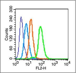
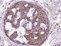
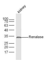
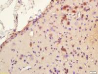
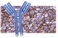
| 产品图片 | 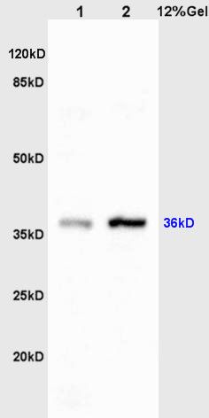 Sample: Liver(Rat)lysate at 30ug; Brain(Rat) lysates at 30ug; Primary: Anti-C-jun/AP-1 (bs-0670R) at 1:200; Secondary: HRP conjugated Goat-Anti-Rabbit IgG(bs-0295G-HRP) at 1: 3000; Predicted band size : 36kD Observed band size : 36kD 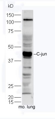 Sample: Lung(Mouse) Lysate at 30ug; Primary: Anti-C-jun (bs-0670R) at 1:300 dilution; Secondary: HRP conjugated Goat-Anti-rabbit IgG(bs-0295G-HRP) at 1:5000; Predicted band size:36 kD Observed band size:40 kD 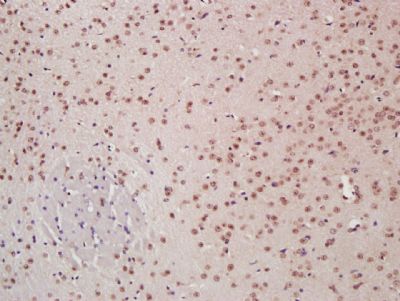 Paraformaldehyde-fixed, paraffin embedded (Mouse brain); Antigen retrieval by boiling in sodium citrate buffer (pH6.0) for 15min; Block endogenous peroxidase by 3% hydrogen peroxide for 20 minutes; Blocking buffer (normal goat serum) at 37°C for 30min; Antibody incubation with (C-jun) Polyclonal Antibody, Unconjugated (bs-0670R) at 1:400 overnight at 4°C, followed by a conjugated secondary antibody (sp-0023) for 20 minutes and DAB staining. 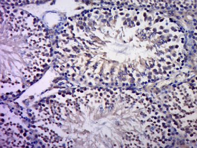 Paraformaldehyde-fixed, paraffin embedded (Rat testis); Antigen retrieval by boiling in sodium citrate buffer (pH6.0) for 15min; Block endogenous peroxidase by 3% hydrogen peroxide for 20 minutes; Blocking buffer (normal goat serum) at 37°C for 30min; Antibody incubation with (C-jun) Polyclonal Antibody, Unconjugated (bs-0670R) at 1:400 overnight at 4°C, followed by a conjugated secondary (sp-0023) for 20 minutes and DAB staining. 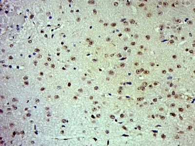 Paraformaldehyde-fixed, paraffin embedded (Mouse brain); Antigen retrieval by boiling in sodium citrate buffer (pH6.0) for 15min; Block endogenous peroxidase by 3% hydrogen peroxide for 20 minutes; Blocking buffer (normal goat serum) at 37°C for 30min; Antibody incubation with (C-jun) Polyclonal Antibody, Unconjugated (bs-0670R) at 1:500 overnight at 4°C, followed by a conjugated secondary (sp-0023) for 20 minutes and DAB staining. 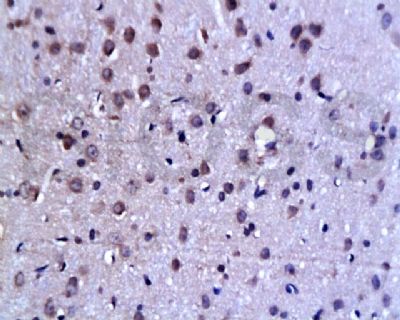 Tissue/cell: rat brain tissue; 4% Paraformaldehyde-fixed and paraffin-embedded; Antigen retrieval: citrate buffer ( 0.01M, pH 6.0 ), Boiling bathing for 15min; Block endogenous peroxidase by 3% Hydrogen peroxide for 30min; Blocking buffer (normal goat serum,C-0005) at 37℃ for 20 min; Incubation: Anti-C-Jun Polyclonal Antibody, Unconjugated(bs-0670R) 1:200, overnight at 4°C, followed by conjugation to the secondary antibody(SP-0023) and DAB(C-0010) staining   Blank control (blue line): HepG2 (blue). Primary Antibody (green line): Rabbit Anti-C-jun antibody (bs-4601R) Dilution: 1μg /10^6 cells; Isotype Control Antibody (orange line): Rabbit IgG . Secondary Antibody (white blue line): Goat anti-rabbit IgG-PE Dilution: 1μg /test. Protocol The cells were fixed with 70% methanol (Overnight at 4℃) and then permeabilized with 90% ice-cold methanol for 20 min at -20℃. Cells stained with Primary Antibody for 30 min at room temperature. The cells were then incubated in 1 X PBS/2%BSA/10% goat serum to block non-specific protein-protein interactions followed by the antibody for 15 min at room temperature. The secondary antibody used for 40 min at room temperature. Acquisition of 20,000 events was performed. |
风险提示:丁香通仅作为第三方平台,为商家信息发布提供平台空间。用户咨询产品时请注意保护个人信息及财产安全,合理判断,谨慎选购商品,商家和用户对交易行为负责。对于医疗器械类产品,请先查证核实企业经营资质和医疗器械产品注册证情况。
 文献和实验
文献和实验乙型肝炎、和HBsAg阳性的患者血清中前S1蛋白,前S1抗原阴转愈早,AHB患者的疗程愈短,预后也愈好。说明前S1抗原及其抗体的检测是急性乙型肝炎的临床诊断,疗效观察和判数据预后的良好指标。 前S1蛋白的分子生物学特性 1.乙肝病毒(HBV)的基因结构 HBV为嗜肝DNA病毒,由一个不完全双链DNA组成,约3200个氨基酸。长链L含4个开放读码框架,为病毒蛋白的编码区:S区、C区、P区、X区。短链S相当于长链的50%-100%,其不固定端可被内原性DNA多聚酶延长,使病毒成为完整
克隆的前癌基因,其转第活性已被证明。 c-myb 禽类成髓细胞增多症病毒辣-转化基因,c-myb的正常细胞性同源物,表达一种细胞核DNA结合蛋白质,被认为调节造血体系统的细胞增生和分化。 c-myc 一种前癌基因,在调节DNA合成、细胞凋亡、分化及细胞周期的进行中起重要作用。c-Myc蛋白质是一种短寿的细胞核磷酸蛋白质,通过联系转录机器和E-合盒无件在转录中起直接作用。c-Myc是一种大性螺旋-环-螺旋亮氨酸的拉链蛋白,相似于USFEY E2F蛋白质家族
纤维素薄膜)上,固相载体以非共价键形式吸附蛋白质,且能保持电泳分离的多肽类型及其生物学活性不变。以固相载体上的蛋白质或多肽作为抗原,与对应的抗 体起免疫反应,再与酶或同位素标记的第二抗体起反应,经过底物显色或放射自显影以检测电泳分离的特异性目的基因表达的蛋白成分。该技术也广泛应用于检测蛋 白水平的表达。二、分类western显色的方法主要有以下几种:i. 放射自显影ii. 底物化学发光ECLiii. 底物荧光ECFiv. 底物DAB呈色现 常用的有底物化学发光ECL和底物DAB呈色,体同水平和实验
 技术资料
技术资料暂无技术资料 索取技术资料

