


- ¥9500
- cytoskeleton
- BK003
- 2025年12月11日

企业认证
相关产品推荐更多 >


万千商家帮你免费找货
0 人在求购买到急需产品
- 详细信息
- 文献和实验
- 技术资料
- 供应商:
研卉生物
- 规格:
30-100 assays
货号:
Product Uses Include
- To show quantitative / qualitative effects on actin polymerization by the addition of a tissue extract, an actin binding protein or compound.
- To show quantitative / qualitative effects on actin polymerization by addition of an F-actin nucleating protein, compound or extract.
- To show quantitative / qualitative effects on steady-state F-actin levels by addition of an F-actin severing protein, compound or tissue extract.
- To show quantitative / qualitative effects on actin depolymerization by addition of an actin binding protein, compound or tissue extract.
Introduction
The Actin Polymerization Biochem Kit™ is based on the enhanced fluorescence of pyrene conjugated actin that occurs during polymerization. The enhanced fluorescence that occurs when pyrene G-actin (monomer) forms pyrene F-actin can be measured in a fluorimeter to follow polymerization over time. Also, by using preformed pyrene F-actin, it is possible to follow depolymerization. Both cell/tissue extracts and purified proteins can be added to the reaction mixture to identify their effect on actin polymerization. The components of the kit can also be used separately for other actin based assays such as a spin-down assays to detect F-actin binding proteins (see also BK001) or size exclusion chromatography to identify G-actin binding proteins. See the About Actin page for more information on assays testing actin binding proteins.
While this kit comes with pyrene labeled skeletal muscle actin, it can also be used to study polymerization of other types of actin such as non-muscle actin (Cat. # APHL99) or cardiac actin (Cat. # AD99). Polymerization assays with these actins can be performed using a 10:1 ratio between the actin you want to study and the included pyrene actin
Kit contents
The kit contains enough materials for 30-100 assays depending on assay volume. The following reagents are included:
- 5 x 1 mg Pyrene labeled actin (Cat. # AP05).
- General Actin Buffer (Cat. # BSA01).
- Actin Polymerization Buffer (Cat. # BSA02).
- ATP 100mM (Cat. # BSA04).
- Tris-HCl pH 7.5, 100 mM
- Manual with detailed protocols and extensive troubleshooting guide.
Equipment needed
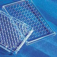
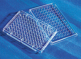
- Fluorescence spectrophotometer (cuvette or 96-well plate) with 4-10 nm bandwidth at 365 nm excitation wavelength, and 4-10 nm bandwidth at 407 nm emission wavelength.
- Small capacity (100-1000 µl) fluorescence spectrophotometer cuvette or 96-well plate.
Example results
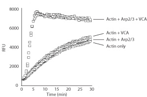
The Actin Polymerization Biochem Kit™ was used to study the effects of Arp2/3 (Cat. # RP01) and the VCA domain of WASP (Cat. # VCG03) on actin polymerization rates. The Arp2/3 complex is an actin filament nucleator but has low nucleating/polymerizing activity on its own. The VCA domain of WASP is an activator of the Arp2/3 complex. Hence, when the Arp2/3 complex is mixed with the WASP VCA domain, these two exert a potent actin polymerizing activity (Fig. 1).
|
||
|
|
|
风险提示:丁香通仅作为第三方平台,为商家信息发布提供平台空间。用户咨询产品时请注意保护个人信息及财产安全,合理判断,谨慎选购商品,商家和用户对交易行为负责。对于医疗器械类产品,请先查证核实企业经营资质和医疗器械产品注册证情况。
 文献和实验
文献和实验Material 1. Source: rabbit skeletal muscle 2. Equipment: • mincer • glass-Teflon-homogenizer with pestle • sephacryl S-300 column (5 x 100 cm) • electrophoretic gels • centrifuge • sterile cheesecloth
Protein purification: skeletal muscle myosin
Overview SKELETAL MUSCLE MYOSIN A two-headed hexomeric actin-activated ATPase consisting of two heavy chains of ~200 kDa and four light chains of ~20kDa. The tail portion
Functional Characterization of Proteins Regulating Actin Assembly
Protocol 7: Measurements of the Treadmilling of Actin Filaments Support Protocol 1: Actin Purification from Rabbit Muscle Support Protocol 2: Preparation of Pyrenyl‐Labeled Actin Support
 技术资料
技术资料暂无技术资料 索取技术资料

