

- ¥1580
- BOISS/雅吉
- 北京
- YS-0914R
- 2025年07月16日
- WB, IHC-P, IF/ICC, P-ELISA
- Rabbit

相关产品推荐更多 >

万千商家帮你免费找货
0 人在求购买到急需产品
- 详细信息
- 文献和实验
- 技术资料
- 免疫原:
Synthetic MAP peptide derived from human beta-Actin
- 亚型:
IgG
- 克隆性:
多克隆
- 宿主:
Rabbit
- 应用范围:
WB, IHC-P, IF/ICC, P-ELISA
- 浓度:
1mg/1ml
- 抗体英文名:
Angiotensin II type 1A receptor
- 规格:
100ul
中文名称血管紧张素Ⅱ1A型受体抗体
别 名AGTR1; Agtr1a; AT1; AT1A; AT1AR; Type 1 angiotensin II receptor; AGTR1_HUMAN; AGTR1B; AT2R1; AT2R1B.
说 明 书50ul 100ul 200ul
研究领域心血管 细胞生物 免疫学
抗体来源Rabbit
克隆类型Polyclonal
交叉反应 Human, Mouse, Rat,
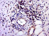
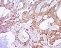
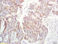
血管紧张素Ⅱ1A型受体抗体产品应用WB=1:500-2000 ELISA=1:500-1000 Flow-Cyt=3ug/Test (石蜡切片需做抗原修复)
not yet tested in other applications.
optimal dilutions/concentrations should be determined by the end user.
分 子 量41kDa
细胞定位细胞膜
性 状Lyophilized or Liquid
浓 度1mg/ml
免 疫 原KLH conjugated synthetic peptide derived from the middle of human Angiotensin II type 1A receptor:101-200/359 <Extracellular>
亚 型IgG
纯化方法affinity purified by Protein A
储 存 液0.01M TBS(pH7.4) with 1% BSA, 0.03% Proclin300 and 50% Glycerol.
保存条件Store at -20 °C for one year. Avoid repeated freeze/thaw cycles. The lyophilized antibody is stable at room temperature for at least one month and for greater than a year when kept at -20°C. When reconstituted in sterile pH 7.4 0.01M PBS or diluent of antibody the antibody is stable for at least two weeks at 2-4 °C.
PubMedPubMed
产品介绍background:
The Angiotensin II type 1 receptor (AT1) is the primary effector of Angiotensin II, a key regulator of blood pressure and fluid homeostasis. It is involved in pathogenesis of several cardiovascular diseases such as hypertension, cardiac hypertrophy and congestive heart failure. Angiotensin II interacts with two types of G-protein coupled membrane receptors, AT1 (type 1) and AT2 (type 2). AT1 has three isoforms in rodents: AT1A (359 aa), AT1B (359 aa), and AT1C (177 aa). Rat AT1's are predicted to contain seven transmembrane domains. The N-terminus is predicted to be extracellular, while the C-terminus is predicted to be cytoplasmic. AT1's are expressed in the liver, kidney, aorta, lung, uterus, ovary, spleen, heart, adrenal and vascular smooth muscle.
Function:
Receptor for angiotensin II. Mediates its action by association with G proteins that activate a phosphatidylinositol-calcium second messenger system.
Subunit:
Interacts with MAS1 (Probable). Interacts with ARRB1.
Subcellular Location:
Cell membrane; Multi-pass membrane protein.
Tissue Specificity:
Liver, lung, adrenal and adrenocortical adenomas.
Post-translational modifications:
C-terminal Ser or Thr residues may be phosphorylated.
DISEASE:
Renal tubular dysgenesis (RTD) [MIM:267430]: Autosomal recessive severe disorder of renal tubular development characterized by persistent fetal anuria and perinatal death, probably due to pulmonary hypoplasia from early-onset oligohydramnios (the Potter phenotype). Note=The disease is caused by mutations affecting the gene represented in this entry.
Similarity:
Belongs to the G-protein coupled receptor 1 family.
SWISS:
P30556
Gene ID:
185
Database links:
Entrez Gene: 185 Human
Entrez Gene: 24180 Rat
Entrez Gene: 81638 Rat
Omim: 106165 Human
SwissProt: P30556 Human
SwissProt: P34976 Rabbit
SwissProt: P25095 Rat
SwissProt: P29089 Rat
Unigene: 477887 Human
Unigene: 728754 Human
Important Note:
This product as supplied is intended for research use only, not for use in human, therapeutic or diagnostic applications.

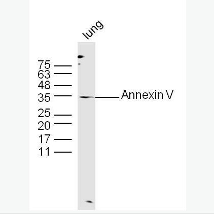
Sample:
Liver (Mouse) Lysate at 40 ug
Lung (Mouse) Lysate at 40 ug
Adrenal gland (Rat) Lysate at 40 ug
Primary: Anti-Angiotensin II type 1A receptor (bs-2132R) at 1/1000 dilution
Secondary: IRDye800CW Goat Anti-Rabbit IgG at 1/20000 dilution
Predicted band size: 41 kD
Observed band size: 56 kD
风险提示:丁香通仅作为第三方平台,为商家信息发布提供平台空间。用户咨询产品时请注意保护个人信息及财产安全,合理判断,谨慎选购商品,商家和用户对交易行为负责。对于医疗器械类产品,请先查证核实企业经营资质和医疗器械产品注册证情况。
 文献和实验
文献和实验反应为V型超敏反应,但多数人认为它是Ⅱ型超敏反应的一种特殊表现形式医|学教育网搜集整理。 (2)重症肌无力:是抗受体抗体介导的功能受抑制的病症。80%以上患者有针对神经肌肉接头处突触后膜上乙酰胆碱受体的抗体,补体参与发病过程。神经肌肉传导障碍导致晨轻暮重、活动后加重、休息可减轻的渐进性骨髓无力及各种受累器官的症状。因受体内吞和在胞内的降解,受体数目减少。 (3)胰岛素抗性糖尿病:有些对胰岛素无反应的糖尿病人抗胰岛素体的自身抗体,受体与自身抗体结合后,胰岛素不能与其受体结合。
底物,即血管紧张素原,在肾素的作用下水解,产生一个十肽,为血管紧张素I。在血浆和组织中,特别是在肺循环血管内皮表面,存在有血管紧张素转换酶,在后者的作用下,血管紧张素I水解,产生一个八肽,为血管紧张素Ⅱ。血管紧张素Ⅱ在血浆和组织中的血管紧张素酶A的作用下,再失去一个氨基酸,成为七肽血管紧张素Ⅲ。上述过程可由图4-28表示。血管紧张素Ⅱ和血管紧张素Ⅲ作用于血管平滑肌和肾上腺皮质等细胞的血管紧张素受体,引起相应的生理效应。 当各种原因引起肾血流灌注减少时,肾素分泌就会
佚名 已有大量的资料证明, 自身免疫 病理损伤是由 自身免疫 应答的产物包括自身抗体和(或)自身致敏淋巴细胞引起的,后者造成病理损伤的机制与各型超敏反应相同(表16-5),以Ⅱ型至v型多见。Ⅱ型超敏反应中,自身抗体与细胞膜或基底膜自身抗原结合,在膜表面形成免疫复合物。后者通过结合并激活补体链锁反应
 技术资料
技术资料暂无技术资料 索取技术资料

