

- ¥1700 - 4000
- GeneTex

- 美国
- GTX130887
- 2025年07月16日
- WB, ICC/IF, IHC-P, IP
- Rabbit
- Human, Mouse, Rat

企业认证
相关产品推荐更多 >

![CD34 antibody [HPCA1/1171]](https://img1.dxycdn.com/2022/0328/634/2953353827299600453-14.jpg!wh200)

万千商家帮你免费找货
0 人在求购买到急需产品
- 详细信息
- 文献和实验
- 技术资料
- 免疫原:
Recombinant protein encompassing a sequence within the center region of human PYK2. The exact sequence is proprietary.
- 亚型:
IgG
- 形态:
Liquid
- 保存条件:
Store as concentrated solution. Centrifuge briefly prior to opening vial. For short-term storage (1-2 weeks), store at 4ºC. For long-term storage, aliquot and store at -20ºC or below. Avoid multiple freeze-thaw cycles.
- 克隆性:
Polyclonal
- 标记物:
Unconjugated
- 适应物种:
Human, Mouse, Rat
- 保质期:
12 months from the shipping date of the product.
- 抗原来源:
Human
- 目录编号:
GTX130887
- 级别:
Primary Antibodies
- 库存:
Available
- 供应商:
GeneTex
- 宿主:
Rabbit
- 应用范围:
WB, ICC/IF, IHC-P, IP
- 浓度:
1.74 mg/ml (Please refer to the vial label for the specific concentration.)
- 靶点:
IP/MS validation was supported by references (PMID:30377418)
- 抗体英文名:
PYK2 antibody
- 抗体名:
PYK2 抗体
- 规格:
100 μl/25 μl
| 规格: | 100 μl | 产品价格: | ¥4000.0 |
|---|---|---|---|
| 规格: | 25 μl | 产品价格: | ¥1700.0 |

Immunoprecipitation of PYK2 protein from A431 whole cell extracts using 5 μg of PYK2 antibody (GTX130887).
Western blot analysis was performed using PYK2 antibody (GTX130887) diluted at 1:500.
EasyBlot anti-Rabbit IgG (GTX221666-01) was used as a secondary reagent.

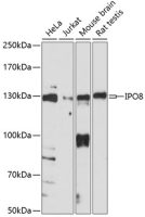
PYK2 antibody detects PYK2 protein by western blot analysis. Various whole cell extracts (30 μg) were separated by 7.5% SDS-PAGE, and the membrane was blotted with PYK2 antibody (GTX130887) diluted at a dilution of 1:1000.

PYK2 antibody detects PYK2 protein at cytoplasm and nucleus in rat brain by immunohistochemical analysis.
Sample: Paraffin-embedded rat brain.
PYK2 antibody (GTX130887) diluted at 1:500.
Antigen Retrieval: Citrate buffer, pH 6.0, 15 min

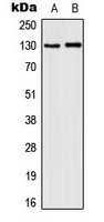
Various tissue extracts (50 μg) were separated by 7.5% SDS-PAGE, and the membrane was blotted with PYK2 antibody (GTX130887) diluted at 1:1000. The HRP-conjugated anti-rabbit IgG antibody (GTX213110-01) was used to detect the primary antibody.
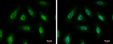
PYK2 antibody detects PYK2 protein at cytoplasm and nucleus by immunofluorescent analysis.
Sample: HeLa cells were fixed in 4% PFA at RT for 15 min.
Green: PYK2 protein stained by PYK2 antibody (GTX130887) diluted at 1:500.
Blue: Hoechst 33342 staining.
Scale bar = 10 μm.

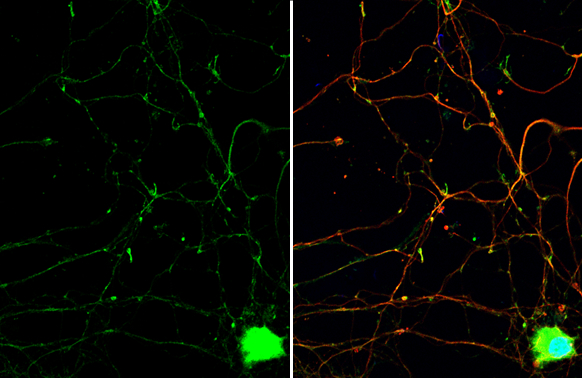
PYK2 antibody detects PYK2 protein by immunofluorescent analysis.
Sample: DIV9 rat E18 primary hippocampal neuron cells were fixed in 4% PFA at RT for 15 min.
Green: PYK2 stained by PYK2 antibody (GTX130887) diluted at 1:500.
Red: beta Tubulin 3/ Tuj1, stained by beta Tubulin 3/ Tuj1 antibody [GT11710] (GTX631836) diluted at 1:500.
Blue: Fluoroshield with DAPI (GTX30920).
风险提示:丁香通仅作为第三方平台,为商家信息发布提供平台空间。用户咨询产品时请注意保护个人信息及财产安全,合理判断,谨慎选购商品,商家和用户对交易行为负责。对于医疗器械类产品,请先查证核实企业经营资质和医疗器械产品注册证情况。
 文献和实验
文献和实验Trenti A et al., Biochem Pharmacol 2018 (PMID:29890142)
Sikorski K et al., Nat Methods 2018 (PMID:30377371)
noworrynoworry 请问各位前辈,Pyk2(Pyk2信号通路)有特异性抑制剂吗?哪里能买到? noworrynoworry 拜托各位,帮帮忙啊 diwen FAK/ Pyk2 tyrosine kinase inhibitor (PF-562271) http://lifesciences.b-bridge.com/products/detail/1440/PF
Generation of Antibody Molecules Through Antibody Engineering
been overcome to a large extent using genetic-engineering techniques to produce chimeric mouse/human and completely human antibodies. Such an approach is particularly suitable because of the domain structure of the antibody molecule ( 2 ), where functional
The importance of antibody molecules was first recognized in the 1890s, when it was shown that immunity to tetanus and diphtheria was caused by antibodies against the bacterial exotoxins (1 ). Around the same time, it was shown that antisera
 技术资料
技术资料暂无技术资料 索取技术资料

