

- ¥1700 - 4000
- GeneTex

- 美国
- GTX127309
- 2025年07月15日
- WB, ICC/IF, IHC-P, IHC-Fr, IP, ChIP assay
- Rabbit
- Human, Mouse, Rat, Rabbit, Bovine

企业认证
相关产品推荐更多 >
![Splunc2 antibody [1F12]](https://img1.dxycdn.com/2022/0328/745/2071730365423500453-14.jpg!wh200)
![NRCAM antibody [7D8C5]](https://img1.dxycdn.com/2022/0328/261/5841871159833500453-14.jpg!wh200)
![CD13 antibody [ER-BMDM1]](https://img1.dxycdn.com/2022/0328/920/0111093443724400453-14.jpg!wh200)
万千商家帮你免费找货
0 人在求购买到急需产品
- 详细信息
- 询价记录
- 文献和实验
- 技术资料
- 免疫原:
Recombinant protein encompassing a sequence within the C-terminus region of human HIF1 alpha. The exact sequence is proprietary.
- 亚型:
IgG
- 形态:
Liquid
- 保存条件:
Store as concentrated solution. Centrifuge briefly prior to opening vial. For short-term storage (1-2 weeks), store at 4ºC. For long-term storage, aliquot and store at -20ºC or below. Avoid multiple freeze-thaw cycles.
- 克隆性:
Polyclonal
- 标记物:
Unconjugated
- 适应物种:
Human, Mouse, Rat, Rabbit, Bovine
- 保质期:
12 months from the shipping date of the product.
- 抗原来源:
Human
- 目录编号:
GTX127309
- 级别:
Primary Antibodies
- 库存:
Available
- 供应商:
GeneTex
- 宿主:
Rabbit
- 应用范围:
WB, ICC/IF, IHC-P, IHC-Fr, IP, ChIP assay
- 浓度:
1.17 mg/ml (Please refer to the vial label for the specific concentration.)
- 靶点:
Knockdown/Knockout validation was supported by references (PMID:25428606)
- 抗体英文名:
HIF1 alpha antibody
- 抗体名:
HIF1 alpha 抗体
- 规格:
100 μl/25 μl
| 规格: | 100 μl | 产品价格: | ¥4000.0 |
|---|---|---|---|
| 规格: | 25 μl | 产品价格: | ¥1700.0 |
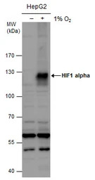
HIF1 alpha antibody detects HIF1 alpha protein by western blot analysis. Un-treated (-) and treated (+, 1% O2 treatment for 24hr) HepG2 whole cell extracts (30 μg) were separated by 7.5% SDS-PAGE, and the membrane was blotted with HIF1 alpha antibody (GTX127309) diluted at 1:1000. The HRP-conjugated anti-rabbit IgG antibody (GTX213110-01) was used to detect the primary antibody.
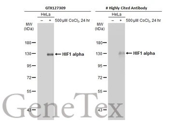
Untreated (–) and treated (+) HeLa whole cell extracts (30 μg) were separated by 7.5% SDS-PAGE, and the membrane was blotted with HIF1 alpha antibody (GTX127309) diluted at 1:1000. The HRP-conjugated anti-rabbit IgG antibody (GTX213110-01) was used to detect the primary antibody.
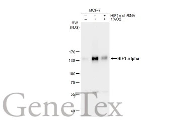
Untreated (–) and treated (+) MCF-7 whole cell extracts (30 μg) were separated by 7.5% SDS-PAGE, and the membrane was blotted with HIF1 alpha antibody (GTX127309) diluted at 1:1000. The HRP-conjugated anti-rabbit IgG antibody (GTX213110-01) was used to detect the primary antibody.
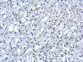
HIF1 alpha antibody detects HIF1 alpha protein at nucleus on human kidney cancer by immunohistochemical analysis.
Sample: Paraffin-embedded human kidney cancer.
HIF1 alpha antibody (GTX127309) dilution: 1:500.
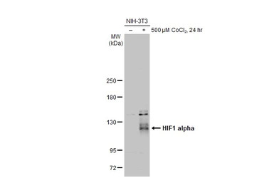
Untreated (–) and treated (+) NIH-3T3 whole cell extracts (30 μg) were separated by 5% SDS-PAGE, and the membrane was blotted with HIF1 alpha antibody (GTX127309) diluted at 1:1000. The HRP-conjugated anti-rabbit IgG antibody (GTX213110-01) was used to detect the primary antibody.
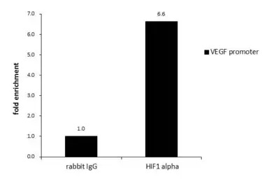
ChIP was performed with HepG2 chromatin extract and 5 μg of either normal rabbit IgG or anti-HIF1 alpha antibody. The precipitated DNA was detected by PCR with primer set targeting to VEGF promoter.
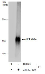
Immunoprecipitation of HIF1 alpha protein from HepG2 whole cell extracts treated with 500 μM CoCl2 for 24 hr using 5 μg of HIF1 alpha antibody (GTX127309).
Western blot analysis was performed using HIF1 alpha antibody (GTX127309) diluted at 1:500.
EasyBlot anti-Rabbit IgG (GTX221666-01) was used as a secondary reagent.
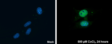
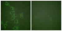
HIF1 alpha antibody detects HIF1 alpha protein at nucleus by immunofluorescent analysis.
Sample: NIH/3T3 cells were fixed in 4% PFA at RT for 15 min.
Green: HIF1 alpha protein stained by HIF1 alpha antibody (GTX127309) diluted at 1:200.
Blue: Hoechst 33342 staining.
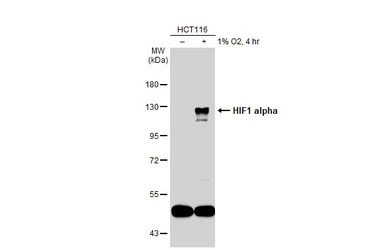
Untreated (–) and treated (+) HCT116 whole cell extracts (30 μg) were separated by 7.5% SDS-PAGE, and the membrane was blotted with HIF1 alpha antibody (GTX127309) diluted at 1:1000. The HRP-conjugated anti-rabbit IgG antibody (GTX213110-01) was used to detect the primary antibody.
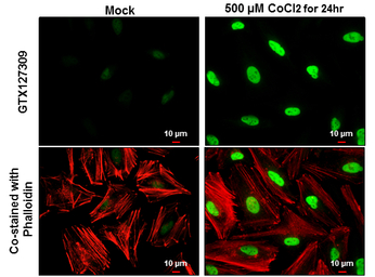
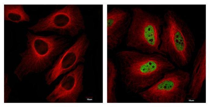
HIF1 alpha antibody detects HIF1 alpha protein at nucleus by immunofluorescent analysis.
Sample: HeLa cells were fixed in 4% PFA at RT for 15 min.
Green: HIF1 alpha protein stained by HIF1 alpha antibody (GTX127309) diluted at 1:500.
Red: Phalloidin, a cytoskeleton marker, diluted at 1:100.
Scale bar = 10 μm.
风险提示:丁香通仅作为第三方平台,为商家信息发布提供平台空间。用户咨询产品时请注意保护个人信息及财产安全,合理判断,谨慎选购商品,商家和用户对交易行为负责。对于医疗器械类产品,请先查证核实企业经营资质和医疗器械产品注册证情况。
- 作者
- 内容
- 询问日期
 文献和实验
文献和实验Sampath Narayanan et al., Commun Biol 2020 (PMID:33318569)
Xiaohu Huang et al., Cancers (Basel) 2021 (PMID:33572152)
Bo Yan et al., Transl Androl Urol 2021 (PMID:34159086)
Chen Zhang et al., Int J Med Sci 2021 (PMID:34522167)
Yi-Chih Tsai et al., Exp Mol Med 2021 (PMID:34702956)
YF Yang et al. et al., J Cell Mol Med 2022 (PMID:35615976)
Giovannucci TA et al., Autophagy 2021 (PMID:34740308)
Shih HJ et al., Sci Rep 2021 (PMID:34824343)
Owada S et al., Anticancer Res 2021 (PMID:34848459)
Peng G et al., J Exp Clin Cancer Res 2021 (PMID:34470658)
Chen YC et al., Cell Biol Toxicol 2021 (PMID:34255241)
N?chel J et al., Mol Cell 2021 (PMID:34245671)
Takenaga K et al., Sci Rep 2021 (PMID:34172808)
Song E et al., bioRxiv 2020 (PMID:32935108)
Ye D et al., Stem Cell Res Ther 2020 (PMID:32894200)
Hsu CG et al., J Immunol 2021 (PMID:34117108)
Sun X et al., Front Genet 2021 (PMID:34220956)
Coronel-Hern?ndez J et al., Front Oncol 2021 (PMID:34123772)
Gampala S et al., Br J Cancer 2021 (PMID:33658640)
Nadeau JR et al., Exp Neurol 2021 (PMID:33684407)
Ezzeddini R et al., J Physiol Biochem 2021 (PMID:33730333)
Nakashima M et al., Cancer Sci 2021 (PMID:33738869)
Kitajima S et al., Sci Rep 2021 (PMID:33633164)
Nishida D et al., Sci Rep 2021 (PMID:33633362)
Weng M et al., Front Cell Dev Biol 2021 (PMID:33665193)
Douiev L et al., Cells 2021 (PMID:33672589)
Chen KC et al., Int J Mol Sci 2020 (PMID:32403414)
Sasaki Y et al., Cancer Sci 2021 (PMID:33393151)
Arellano-Buend?a AS et al., Antioxidants (Basel) 2020 (PMID:33203103)
Cirotti C et al., EMBO Rep 2020 (PMID:33245190)
Eric Song et al., J Exp Med 2021 (PMID:33433624)
Ueshima E et al., Liver Cancer 2020 (PMID:32071910)
Chen HY et al., Chem Biol Interact 2020 (PMID:32980322)
Hou J et al., Nat Cell Biol 2020 (PMID:32929201)
Tazawa K et al., Am J Pathol 2020 (PMID:32919979)
Owada S et al., Anticancer Res 2020 (PMID:32878795)
Singh M et al., Cancer Res 2019 (PMID:30952634)
Zhao Y et al., Am J Physiol Renal Physiol 2020 (PMID:32715763)
Bailey CM et al., Nanomedicine 2020 (PMID:32738299)
Chan CH et al., Aging (Albany NY) 2020 (PMID:32759461)
Takaki H et al., J Vasc Interv Radiol 2020 (PMID:32800663)
Mochizuki M et al., Stem Cell Res Ther 2020 (PMID:32660544)
Lai HH et al., J Clin Invest 2018 (PMID:29251629)
Kuriyama S et al., Cell Death Discov 2018 (PMID:29531808)
Eric Song et al., 2020 (Epub)
Kitajima S et al., Oncotarget 2018 (PMID:29721188)
Dou YQ et al., Theranostics 2020 (PMID:31938060)
Ve?e?a J et al., Stem Cell Res 2020 (PMID:32276221)
Gojkovic M et al., Acta Physiol (Oxf) 2020 (PMID:32129933)
Basaco T et al., Pharmaceuticals (Basel) 2018 (PMID:30487460)
Chen W et al., Oncol Lett 2019 (PMID:30854062)
Kato H et al., Oncogene. 2020 (PMID:32071397)
Manuprasert W et al., Asian Pac J Allergy Immunol 2019 (PMID:31837216)
Chua KV et al., Cells 2019 (PMID:31905895)
Fujii M et al., Int Endod J 2020 (PMID:31910287)
Zhang S et al., Gene 2020 (PMID:31972307)
Wu TH et al., Int J Mol Sci 2019 (PMID:31775307)
Pan CH et al., Aging (Albany NY) 2019 (PMID:31584878)
Zheng J et al., J Ethnopharmacol 2019 (PMID:31654796)
Ho YJ et al., Theranostics 2019 (PMID:31695774)
Li F et al., World J Pediatr 2019 (PMID:31535281)
Loh XY et al., Cancer Res 2019 (PMID:31551365)
Tang K et al., Oncogene 2019 (PMID:31409901)
Kuo YL et al., Sci Rep 2019 (PMID:31311941)
Lai PY et al., Physiol Genomics 2019 (PMID:31373541)
Offer S et al., J Exp Clin Cancer Res 2019 (PMID:31174567)
Holzer T et al., J Cell Biol 2019 (PMID:31085560)
Ma Z et al., Cell Cycle 2019 (PMID:31107137)
Zhou M et al., Sci Rep 2019 (PMID:30988335)
Wang X et al., J Steroid Biochem Mol Biol 2019 (PMID:31028793)
Song S et al., Am J Transl Res 2019 (PMID:31105825)
Ezzeddini R et al., Life Sci 2019 (PMID:30914315)
Kurelac I et al., Nat Commun 2019 (PMID:30796225)
Wenli Chen et al., Oncology Letters 2019
Yasunaga M et al., Cancer Sci 2019 (PMID:30537002)
Hsu HL et al., Int J Mol Sci 2019 (PMID:30609861)
Bin? L et al., Oncotarget 2017 (PMID:29137374)
Nagalakshmi VK et al., Clin Sci (Lond) 2018 (PMID:30442812)
Sun W et al., Sci Rep 2017 (PMID:28775317)
Shih JW et al., Nat Commun 2017 (PMID:28639619)
Kraus RJ et al., PLoS Pathog 2017 (PMID:28617871)
Wang M et al., Theranostics 2017 (PMID:28435451)
Masahiro Yasunaga et al., bioRxiv 2018 (Epub)
Xu G et al., Autophagy 2018 (PMID:30103670)
Di K et al., Oncotarget 2016 (PMID:27764809)
Sasa K et al., Sci Rep 2018 (PMID:30002387)
Fu JL et al., Hum Reprod 2018 (PMID:29982401)
Jou YC et al., Oncotarget 2016 (PMID:27557492)
Hassan A et al., PLoS One 2018 (PMID:29775479)
Yamamura H et al., Am J Physiol Cell Physiol 2018 (PMID:29768048)
Ho SY et al., BMC Cancer 2018 (PMID:29703163)
Demetriades C et al., Nat Commun 2016
In situ hybridisation to alpha satellite sequences (chromosome specific)
Alpha satellite sequences, whilst highly repetitive, are specific to each individual chromosome. These sequences flank the centromeres and can present a target measured in megabases. In this protocol a biotin or digoxigenin labelled DNA
Immunohistochemical Detection of Tumour Hypoxia
In this chapter, we describe the use of immunohistochemical methods to detect hypoxia in tumour tissue sections, utilising antibodies specific for endogenous proteins hypoxia inducible factor 1 alpha (Hif1α) and glucose transporter 1 (Glut
marrow. The prime suspect: an adhesion molecule called alpha2 integrin. The molecule showed increased expression on memory cells, and its ligand is predominantly expressed in bone marrow tissue. Also, when the researchers blocked alpha2 integrin
 技术资料
技术资料暂无技术资料 索取技术资料

