

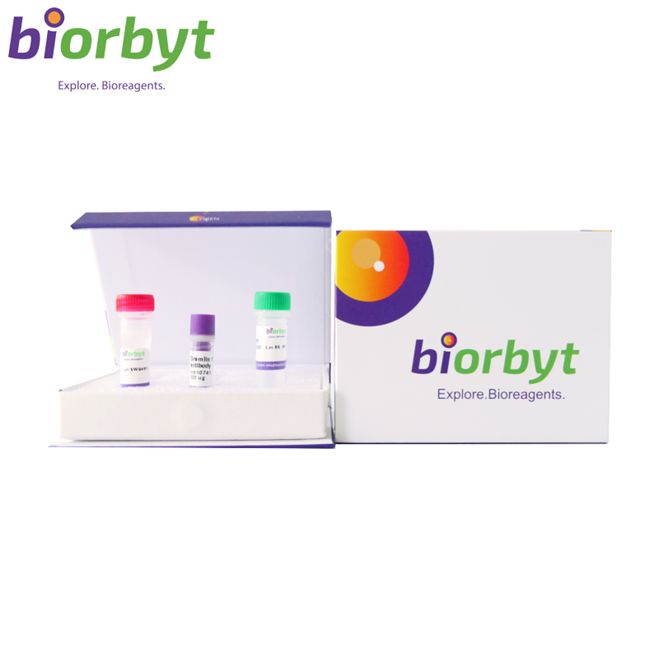
- ¥7371
- Biorbyt

- orb151862
- 英国
- 2026年07月29日
- ICC, IF, IHC, IP, WB
- Rabbit
- Bovine, Canine, Human, Monkey, Mouse, Rabbit, Rat

企业认证
相关产品推荐更多 >
CD142 Rabbit Polyclonal Antibody 抗体,orb339106,Biorbyt
¥1638NF-kappaB p65 Rabbit Polyclonal Antibody 抗体,orb315684,Biorbyt
¥1638
NG2 Recombinant Rabbit Monoclonal Antibody 抗体,orb704325,Biorbyt
¥2223
TLR2 Rabbit Polyclonal Antibody (Biotin) 抗体,orb2099263,Biorbyt
¥0
KRT36 Rabbit Polyclonal Antibody (Biotin) 抗体,orb2096368,Biorbyt
¥0
万千商家帮你免费找货
0 人在求购买到急需产品
相关阅读
源叶生物 十万种现货试剂速发,品质稳、品类全,实验不再等!Takara 秋促来袭,提取+反转录+定量套装仅 1200 元abinScience 体内级抗体 全年锁价 低至 ¥1200阿拉丁 22万+ 科研级产品现货速发,品质严控、科研无忧- 详细信息
- 文献和实验
- 技术资料
- 抗体名:
TNF-R1 Antibody (Biotin) 抗体
- 抗体英文名:
TNF-R1 Antibody (Biotin)
- 靶点:
TNF-R1
- 浓度:
1 mg/ml
- 应用范围:
ICC, IF, IHC, IP, WB
- 宿主:
Rabbit
- 适应物种:
Bovine, Canine, Human, Monkey, Mouse, Rabbit, Rat
- 保质期:
12 months from date of receipt.
- 抗原来源:
详询
- 目录编号:
orb151862
- 级别:
科研
- 库存:
99
- 供应商:
Biorbyt
- 标记物:
Biotin
- 克隆性:
Polyclonal
- 保存条件:
详询
- 免疫原:
详询
- 规格:
100 ug
描述: Rabbit polyclonal antibody against TNFR1 conjugated to Biotin
别名: Tumor necrosis factor receptor 1, TNFR-1, TNFRSF1A, TNFAR, TNFR1
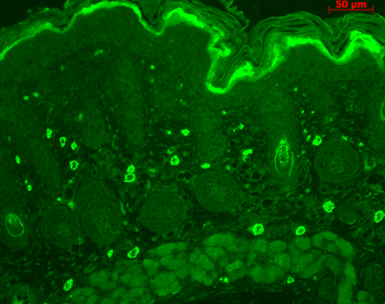
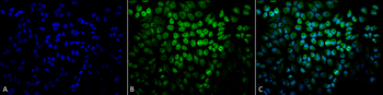
应用稀释比例: WB (1:1000), IHC (1:100), ICC/IF (1:100)
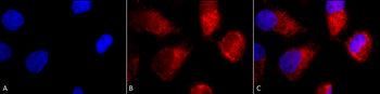
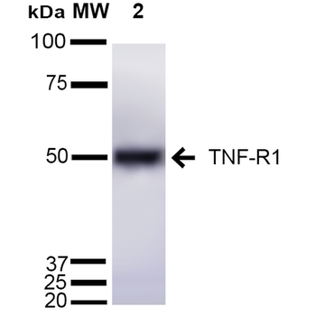
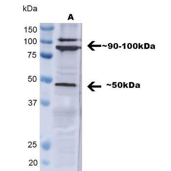
应用说明: 1 µg/ml was sufficient for detection of TNFR1 in 20 µg of Hela lysate by colorimetric immunoblot analysis using Goat anti-rabbit IgG:HRP as the secondary antibody.
免疫原: Peptide corresponding to AA 20-43 of the mouse TNF-R1 sequence, identical to rat and human over those residues
分子量: 55kDa
防腐剂: 136.36mM Ethanolamine, 133.23 mM Chlorides, 9.55mM Phosphates, 9.55mM Sodium Bicarbonate.
纯化方法: Peptide Affinity Purified
特异性: Detects ~55kDa. Other bands present may be the result of oligomerization, self-aggregation and/or cleavage of the TNF-R1 extracellular domain.
研究领域: Cell Biology
UniProt ID: P19438
NCBI 参考序列: P19438
Entrez 基因 ID: 8666
保存条件: Conjugated antibodies should be stored according to the product label
备注: For research use only.
风险提示:丁香通仅作为第三方平台,为商家信息发布提供平台空间。用户咨询产品时请注意保护个人信息及财产安全,合理判断,谨慎选购商品,商家和用户对交易行为负责。对于医疗器械类产品,请先查证核实企业经营资质和医疗器械产品注册证情况。
 文献和实验
The Avidin-Biotin Complex (ABC) Method and Other Avidin-Biotin Binding Methods
These methods involve, at their core, the vitamin biotin and the protein avidin, which bind together irreversibly. By establishing a biotin link, through avidin, between the horseradish peroxidase enzyme and a secondary antibody reagent
Tumor necrosis factor-α (TNF-α) is a cytokine (CK) that possesses a wide variety of biological activities, including potent antitumor activities (1 ) and immunomodulatory properties mediated through its binding to two TNF receptors (p55
The Avidin-Biotin Complex(ABC) Method
, between the horseradish peroxidase enzyme and a secondary antibody reagent, enzyme localization can be achieved at the site of primary antibody interaction with the specimen (see ) Fig. 1 ). The biotin molecule is small and can be easily conjugated to immunoglobulin
 技术资料
暂无技术资料 索取技术资料