

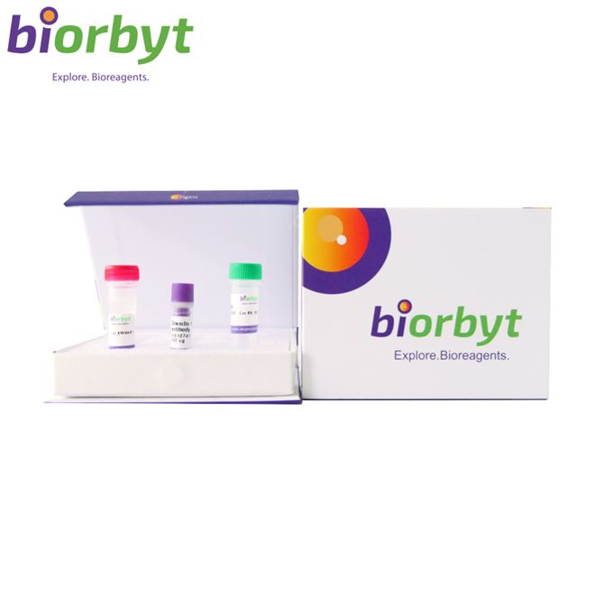
- ¥6903
- Biorbyt

- orb151496
- 英国
- 2026年07月29日
- FC, ICC, IF, IHC, WB
- Rabbit
- Bovine, Drosophila, Frog, Gallus, Human, Mouse, Rat, Sheep

企业认证
相关产品推荐更多 >
IRS1 Rabbit Polyclonal Antibody 抗体,orb315570,Biorbyt
¥1638
ROBO1 Antibody 抗体,orb345475,Biorbyt
¥10413
PPEF2 Rabbit Polyclonal Antibody (FITC) 抗体,orb2089548,Biorbyt
¥0Histone H2A.Z (Acetyl-K7) Rabbit Polyclonal Antibody 抗体,orb334672,Biorbyt
¥1872
Phospho-Hsp90 beta (Ser226) Rabbit Polyclonal Antibody (PerCP-Cy5.5) 抗体,orb1589936,Biorbyt
¥6201
万千商家帮你免费找货
0 人在求购买到急需产品
相关阅读
源叶生物 十万种现货试剂速发,品质稳、品类全,实验不再等!Takara 秋促来袭,提取+反转录+定量套装仅 1200 元abinScience 体内级抗体 全年锁价 低至 ¥1200阿拉丁 22万+ 科研级产品现货速发,品质严控、科研无忧- 详细信息
- 文献和实验
- 技术资料
- 抗体名:
Erk1/2 Antibody (Biotin) 抗体
- 抗体英文名:
Erk1/2 Antibody (Biotin)
- 靶点:
Erk1/2
- 浓度:
1 mg/ml
- 应用范围:
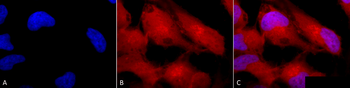
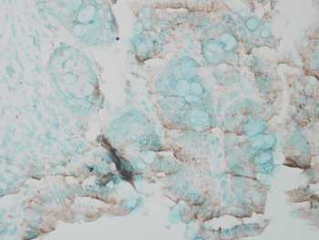
FC, ICC, IF, IHC, WB
- 宿主:
Rabbit
- 适应物种:
Bovine, Drosophila, Frog, Gallus, Human, Mouse, Rat, Sheep
- 保质期:
12 months from date of receipt.
- 抗原来源:
详询
- 目录编号:
orb151496
- 级别:
科研
- 库存:
99
- 供应商:
Biorbyt
- 标记物:
Biotin
- 克隆性:
Polyclonal
- 保存条件:
详询
- 免疫原:
详询
- 规格:
100 ul
描述: Rabbit polyclonal to Erk1 (Biotin). The extracellular signal-regulated kinases 1 and 2 (ERK1 and ERK2), also called p44 and p42 MAP kinases, are members of the Mitogen Activated Protein Kinase (MAPK) family of proteins found in all eukaryotes. Because the 44 kDa ERK1 and the 42 kDa ERK2 are highly homologous and both function in the same protein kinase cascade, the two proteins are often referred to collectively as ERK1/2 or p44/p42 MAP kinase. They are both located in the cytosol and mitochondria. While the role of cytosol ERK1/2 is well studied and involved in multiple cellular functions, the role of mitochondrial ERK1/2 remains poorly understood. Both ERK 1 and 2 are activated by MEK1 or MEK2, by dual phosphorylation of a threonine and tyrosine residue in the activation loop (TEY motif) (1, 3). Either phosphorylation alone can induce an electrophoretic mobility shift, but both are required for activation of the kinase. This dual phosphorylation is efficiently detected by phosphorylation state-specific antibody directed to the pTEpY motif. Once activated, MAP kinases phosphorylate a broad spectrum of substrates, including cytoskeletal proteins, translation regulators, transcription factors, and the Rsk family of protein kinases. ERK1/2 activation is generally thought to confer a survival advantage to cells; however there is increasing evidence that suggests that the activation of ERK1/2 also contributes to cell death under certain conditions. ERK1/2 also is activated in neuronal and renal epithelial cells upon exposure to oxidative stress and toxicants or deprivation of growth factors, and inhibition of the ERK pathway blocks apoptosis..
别名: ERK1, ERK2, ERT1, ERT2, MAP kinase 1, MAP kinase 2, MAPK1, MAPK2, MAPK3, p38, p40, p41, p41mapk, p42 MAPK, p44 ERK1, p44 MAPK, PRKM1, PRKM2, PRKM3
应用稀释比例: WB (1:1000), IHC (1:100), ICC/IF (1:100), FCM (1:100)
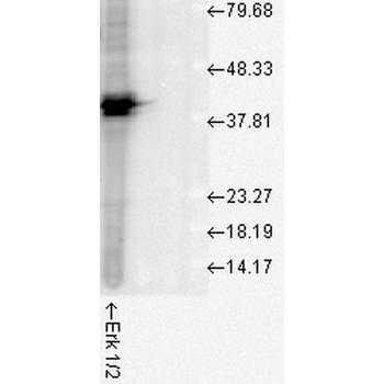
应用说明: A 1:1000 dilution was sufficient for detection of ERK1/2 in 20 µg of HeLa cell lysate by ECL immunoblot analysis.
免疫原: A 35 residue synthetic peptide, corresponding to Erk1 MAP kinase with the CGG spacer group added and the peptide coupled to KLH.
分子量: 42kDa (ERK2).
防腐剂: 136.36mM Ethanolamine, 133.23 mM Chlorides, 9.55mM Phosphates, 9.55mM Sodium Bicarbonate.
纯化方法: Peptide Affinity Purified
特异性: Detects ~44kda (ERK1) and ~42kDa (ERK2).
研究领域: Cell Biology
UniProt ID: P21708
NCBI 参考序列: NP_059043.1
Entrez 基因 ID: 50689
保存条件: Conjugated antibodies should be stored according to the product label
备注: For research use only.
风险提示:丁香通仅作为第三方平台,为商家信息发布提供平台空间。用户咨询产品时请注意保护个人信息及财产安全,合理判断,谨慎选购商品,商家和用户对交易行为负责。对于医疗器械类产品,请先查证核实企业经营资质和医疗器械产品注册证情况。
 文献和实验
The Avidin-Biotin Complex (ABC) Method and Other Avidin-Biotin Binding Methods
These methods involve, at their core, the vitamin biotin and the protein avidin, which bind together irreversibly. By establishing a biotin link, through avidin, between the horseradish peroxidase enzyme and a secondary antibody reagent
The Avidin-Biotin Complex(ABC) Method
, between the horseradish peroxidase enzyme and a secondary antibody reagent, enzyme localization can be achieved at the site of primary antibody interaction with the specimen (see ) Fig. 1 ). The biotin molecule is small and can be easily conjugated to immunoglobulin
sample to 1 mg/ml (or less). Prepare 1 mg/ml solution of NHS-Biotin in DMSO. Add NHS-Biotin in DMSO to antibody at desired ratio e.g. for 1:8 ratio add 125 ml of NHS-Biotin to 1mg of Ab Incubate on rotator for 4 hours at room
 技术资料
暂无技术资料 索取技术资料