

Erk1/2 Antibody 抗体,orb1822436,Biorbyt产品图

Erk1/2 Antibody 抗体,orb1822436,

Biorbyt
收藏
  • ¥6201
  • Biorbyt已认证
  • orb1822436
  • 英国
  • 2026年08月06日
  • FC, ICC, IF, IHC, WB
  • Rabbit
  • Bovine, Drosophila, Frog, Gallus, Human, Mouse, Rat, Sheep

avatar
品牌商
13 年钻石会员
  • 企业认证

    点击 QQ 联系

    • 详细信息
    • 文献和实验
    • 技术资料
    • 抗体名:

      Erk1/2 Antibody 抗体

    • 抗体英文名:

      Erk1/2 Antibody

    • 靶点:

      Erk1/2

    • 浓度:

      1 mg/ml

    • 应用范围:

      FC, ICC, IF, IHC, WB

    • 宿主:

      Rabbit

    • 适应物种:

      Bovine, Drosophila, Frog, Gallus, Human, Mouse, Rat, Sheep

    • 保质期:

      12 months from date of receipt.

    • 抗原来源:

      详询

    • 目录编号:

      orb1822436

    • 级别:

      科研

    • 库存:

      99

    • 供应商:

      Biorbyt

    • 标记物:

      Unconjugated

    • 克隆性:

      Polyclonal

    • 保存条件:

      详询

    • 免疫原:

      详询

    • 规格:

      100 ul

    类别: Antibodies

    描述: Rabbit Anti-Rat Erk1/2 Polyclonal

    别名: ERK1, ERK2, ERT1, ERT2, MAP kinase 1, MAP kinase 2, MAPK1, MAPK2, MAPK3, p38, p40, p41, p41mapk, p42 MAPK, p44 ERK1, p44 MAPK, PRKM1, PRKM2, PRKM3

    应用稀释比例: WB (1:1000), IHC (1:100), ICC/IF (1:100), FCM (1:100); optimal dilutions for assays should be determined by the user.

    应用说明: A 1:1000 dilution was sufficient for detection of ERK1/2 in 20 µg of HeLa cell lysate by ECL immunoblot analysis.

    免疫原: A 35 residue synthetic peptide, corresponding to Rat Erk1 MAP kinase with the CGG spacer group added and the peptide coupled to KLH.

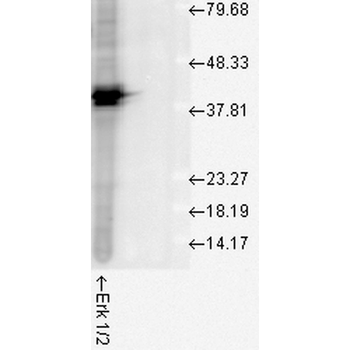
    分子量: 42kDa (ERK2).

    防腐剂: PBS pH 7.4, 50% glycerol, 0.09% sodium azide. Storage buffer changes when conjugated.

    纯化方法: Peptide Affinity Purified

    特异性: Detects ~44kda (ERK1) and ~42kDa (ERK2).

    研究领域: Cell Biology

    UniProt ID: P21708

    NCBI 参考序列: NP_059043.1

    Entrez 基因 ID: 50689

    保存条件: Maintain refrigerated at 2-8°C for up to 2 weeks. For long term storage store at -20°C in small aliquots to prevent freeze-thaw cycles.

    备注: For research use only.
    产品细节图片1
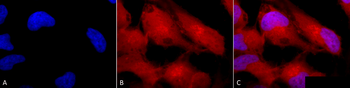
    Immunocytochemistry/Immunofluorescence analysis using Rabbit Anti-Erk1/2 Polyclonal Antibody. Tissue: Cervical cancer cell line (HeLa). Species: Human. Fixation: 2% Formaldehyde for 20 min at RT. Primary Antibody: Rabbit Anti-Erk1/2 Polyclonal Antibody at 1:100 for 12 hours at 4°C. Secondary Antibody: APC Goat Anti-Rabbit (red) at 1:200 for 2 hours at RT. Counterstain: DAPI (blue) nuclear stain at 1:40000 for 2 hours at RT. Localization: Cytoplasm. Nucleus. Magnification: 100x. (A) DAPI (blue) nuclear stain. (B) Anti-Erk1/2 Antibody. (C) Composite.

    产品细节图片2
    Western blot analysis of Human Cell line lysates showing detection of ERK1 protein using Rabbit Anti-ERK1 Polyclonal Antibody. Load: 15 μgprotein. Block: 1.5% BSA for 30 minutes at RT. Primary Antibody: Rabbit Anti-ERK1 Polyclonal Antibody at 1:1000 for 2 hours at RT. Secondary Antibody: Donkey Anti-Rabbit IgG: HRP for 1 hour at RT.

    产品细节图片3
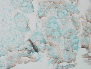
    Immunohistochemistry analysis using Rabbit Anti-ERK1 Polyclonal Antibody. Tissue: Inflamed colon. Species: Mouse. Fixation: Formalin. Primary Antibody: Rabbit Anti-ERK1 Polyclonal Antibody at 1:25000 for 12 hours at 4°C. Secondary Antibody: Biotin Goat Anti-Rabbit at 1:2000 for 1 hour at RT. Counterstain: Methyl Green at 200uL for 2 min at RT.

    产品细节图片4
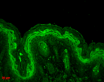
    Immunohistochemistry analysis using Rabbit Anti-ERK1 Polyclonal Antibody. Tissue: backskin. Species: Mouse. Fixation: Bouin's Fixative Solution. Primary Antibody: Rabbit Anti-ERK1 Polyclonal Antibody at 1:100 for 1 hour at RT. Secondary Antibody: FITC Goat Anti-Rabbit (green) at 1:50 for 1 hour at RT. Localization: Cytoplasm.

    产品细节图片5
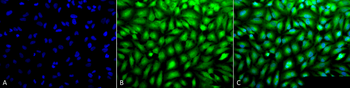
    Immunocytochemistry/Immunofluorescence analysis using Rabbit Anti-Erk1/2 Polyclonal Antibody. Tissue: Cervical cancer cell line (HeLa). Species: Human. Fixation: 2% Formaldehyde for 20 min at RT. Primary Antibody: Rabbit Anti-Erk1/2 Polyclonal Antibody at 1:100 for 12 hours at 4°C. Secondary Antibody: FITC Goat Anti-Rabbit (green) at 1:200 for 2 hours at RT. Counterstain: DAPI (blue) nuclear stain at 1:40000 for 2 hours at RT. Localization: Cytoplasm. Nucleus. Magnification: 20x. (A) DAPI (blue) nuclear stain. (B) Anti-Erk1/2 Antibody. (C) Composite.

    产品细节图片6
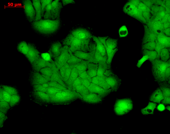
    Immunocytochemistry/Immunofluorescence analysis using Rabbit Anti-ERK1 Polyclonal Antibody. Tissue: HaCaT cells. Species: Human. Fixation: Cold 100% methanol at -20°C for 10 minutes. Primary Antibody: Rabbit Anti-ERK1 Polyclonal Antibody at 1:100 for 12 hours at 4°C. Secondary Antibody: FITC Goat Anti-Rabbit at 1:50 for 1-2 hours at RT in dark. Localization: Cytoplasm. Nucleus.

    风险提示:丁香通仅作为第三方平台,为商家信息发布提供平台空间。用户咨询产品时请注意保护个人信息及财产安全,合理判断,谨慎选购商品,商家和用户对交易行为负责。对于医疗器械类产品,请先查证核实企业经营资质和医疗器械产品注册证情况。

    图标文献和实验
    相关实验
    • secondary antibody review -- data from 99 publications

      cytometry   used as a control to detect cell responses targeted antigen   7       Alexa Fluor 488         7       Cy3         8 goat IgG   Alexa Fluor 488   1:2000 detect antibody binding in human embryonic kidney 293T cells Invitrogen 9 donkey

    • L-02细胞系特性

      secondary antibody were purchased from GIBCO-BRL. Phosphospecific rabbit polyclonal antibodies against 180Thr and 182Tyr dual-phosphorylated p38, against total p38 (phosphorylation state independent), against 204Tyr-phosphorylated ERK1/2 and against total

    • Stripping Western Blots

      per immunoprecipitation. You can use less (as little as 50) but the higher amount gives you a good, easily detectable amount of activity. 9.) To the lysate, add 1.5 μg of anti-ERK antibody (Santa Cruz, SC154 (Erk2) or SC93 (Erk1)), and incubate, with rocking

    图标技术资料

    暂无技术资料 索取技术资料

    同类产品报价

    产品名称
    产品价格
    公司名称
    报价日期
    ¥2106
    武汉博欧特生物科技有限公司
    2026年08月07日询价
    ¥1800
    爱必信(上海)生物科技有限公司
    2025年07月05日询价
    询价
    StressMarq Biosciences Inc.
    2026年09月25日询价
    询价
    上海优宁维生物科技股份有限公司
    2026年06月21日询价
    ¥1524.60
    深圳欣博盛生物科技有限公司
    2025年07月16日询价
    Erk1/2 Antibody 抗体,orb1822436,Biorbyt
    ¥6201