

- 询价
- BIO-RAD

- MCA1266SBB675
- 2025年12月27日
- F
- Mouse

企业认证
相关产品推荐更多 >

万千商家帮你免费找货
0 人在求购买到急需产品
- 详细信息
- 文献和实验
- 技术资料
- 免疫原:
Spleen and bone marrow cells from CE mice.
- 亚型:
IgG2a
- 形态:
Purified IgG conjugated to StarBright Blue 675 - liquid
- 克隆性:
Monoclonal Antibody
- 标记物:
StarBright Blue 675
- 适应物种:
Mouse
- 保质期:
自发货之日起 12 个月
- 目录编号:
PK136
- 供应商:
伯乐生命医学产品(上海)有限公司
- 应用范围:
F
- 浓度:
For information on the concentration of our StarBright Dye conjugated reagents please visit our FAQ page.
- 抗体名:
CD161 / NK1.1 antibody | PK136
- 规格:
100 Tests/0.5ml
小鼠抗小鼠 CD161 / NK1.1 抗体(克隆号 PK136)可识别小鼠 NK1.1 细胞表面抗原,该抗原是由 NKR-P1 基因家族成员编码的一种细胞表面糖蛋白。NK1.1 表面抗原也被称为 CD161b / CD161c 和 Ly-55。
在小鼠中,NKR-P1 家族有三个成员:NKR-P1A、-B 和 -C,而在人类中仅发现一个成员。人类蛋白被命名为 CD161,小鼠蛋白则被称为 CD161a、-b、-c 等。
尽管此前认为 PK136 抗体仅识别 CD161c,但近期数据显示,它也可能与 CD161b 发生反应。CD161c 本身的表达在小鼠中具有品系特异性,但 PK136 对 CD161b 的识别则更为复杂——因为该抗体只能标记部分 CD161b 阳性品系。据报道,CD161c 的结合对 NK 细胞具有激活功能,而 CD161b 的结合则具有抑制作用。
小鼠抗小鼠 NK1.1 抗原抗体(克隆号 PK136)可用于鉴定特定品系小鼠中的 NK 细胞(在 C57BL、FVB/N 和 NZB 品系中呈阳性,在 AKR 和 BALB/c 中呈阴性),也表达于少数 T 细胞和单核细胞亚群。该抗体还已用于体内 NK 细胞的清除(Wang et al. 2022)以及体外 NK 细胞的活化(Kung and Miller 1995)。
| 项目 | 内容 |
|---|---|
| 靶物种 | 小鼠 |
| 物种交叉反应 | 靶物种:大鼠、人 交叉反应性:以上均无反应(✗) 注意:抗体反应性及工作条件可能因物种而异。 |
| 产品形式 | 纯化IgG,与StarBright Blue 675荧光染料偶联,液体 |
| 制备方法 | 通过Protein A亲和层析从组织培养上清液中纯化获得IgG |
| 缓冲溶液 | 磷酸盐缓冲盐水 |
| 防腐剂/稳定剂 | 0.09% NaN₃ 1% 牛血清白蛋白 0.1% Pluronic F68 0.1% PEG 3350 0.05% Tween 20 |
| 免疫原 | 来自CE小鼠的脾脏与骨髓细胞 |
| 蛋白浓度 | 请访问厂家FAQ页面查询StarBright染料偶联试剂的浓度信息 |
| 融合伴侣 | 免疫 (C3H × BALB/c) F1杂合小鼠脾细胞 + Sp2/0-Ag14骨髓瘤细胞系 |
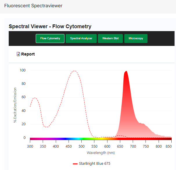
| 光谱特性 | 荧光染料:StarBright Blue 675 最大激发波长:476 nm 最大发射波长:675 nm |
| 用途说明 | 仅供研究使用 |
| 保质期 | 自发货之日起12个月 |
| 专利信息 | 受美国专利号10,150,841及相关国际专利保护。 |
储存信息
本品于环境温度运输。
请于 +4°C 条件下储存。
请勿冷冻。
本品应原液保存,请勿稀释。
应用说明
据报道,本品适用于以下应用。相关信息来自我们实验室的内部测试、同行评审文献或原始研发人员的个人交流。具体信息请参阅相关参考文献。如需一般实验方案建议,请访问抗体实验方案页面。
| 应用名称 | 已验证 | 推荐最低稀释度 | 推荐最高稀释度 |
|---|---|---|---|
| 流式细胞术 | ✓ | 原液 |
若本品未针对某项特定技术进行测试,并不代表其不适用于该技术。推荐的参考工作稀释度仅供参考,建议用户在其体系中通过适当的阴性/阳性对照,自行滴定确定最佳使用浓度。
流式细胞术
建议使用 5 µL 工作稀释液标记 100 µL 体积中的 106 个细胞。最佳实验流程建议在上样前以 6,000 g 离心 5 分钟。
In the mouse the NKR-P1 family has three members, NKR-P1A, -B and -C, whilst in the human only one member has been identified. The human protein has received the designation CD161, and the mouse proteins have been referred to as CD161a, -b, -c etc.
Although previously thought to recognize only CD161c, recent data has shown that the PK136 antibody may also react with CD161b. CD161c expression itself is strain specific in mice, but recognition of CD161b by PK136 appears to be even more complex, as only some CD161b positive strains are labelled by the antibody. Engagement of CD161c has been reported to have activating function in NK cells, whilst engagement of CD161b is inhibitory.
Mouse anti Mouse NK1.1 Antigen antibody, clone PK136 is useful for the identification of NK cells in selected strains of mice (positive on C57BL, FVB/N and NZB, but negative on AKR and BALB/c) and is also expressed by rare subsets of T cells and monocytes. Mouse anti Mouse NK1.1 antibody, clone PK136 has also been used for in vivo depletion of NK cells (Wang et al. 2022) and in vitro activation of NK cells (Kung and Miller 1995).
储存信息
Store at +4°C.
DO NOT FREEZE.
This product should be stored undiluted.
应用
| Application Name | Verified | Min Dilution | Max Dilution |
|---|---|---|---|
| Flow Cytometry | ✓ | Neat |
Flow Cytometry
Use 5μl of the suggested working dilution to label 106 cells in 100μl. Best practices suggest a 5 minutes centrifugation at 6,000g prior to sample application.
风险提示:丁香通仅作为第三方平台,为商家信息发布提供平台空间。用户咨询产品时请注意保护个人信息及财产安全,合理判断,谨慎选购商品,商家和用户对交易行为负责。对于医疗器械类产品,请先查证核实企业经营资质和医疗器械产品注册证情况。
 文献和实验
文献和实验Source Reference
- Koo, G.C. & Peppard, J.R. (1984) Establishment of monoclonal anti-Nk-1.1 antibody.
Hybridoma. 3 (3): 301-3.
References for CD161 / NK1.1 antibody
- Koo, G.C. et al. (1986) The NK-1.1(-) mouse: a model to study differentiation of murine NK cells.
J Immunol. 137 (12): 3742-7. - Kung, S.K. & Miller RG (1995) The NK1.1 antigen in NK-mediated F1 antiparent killing in vitro.
J Immunol. 154 (4): 1624-33. - Wang, M. et al. (1998) Natural killer cell depletion fails to influence initial CD4 T cell commitment in vivo in exogenous antigen-stimulated cytokine and antibody responses.
J Immunol. 160 (3): 1098-105. - Halin, C. et al. (2002) Enhancement of the antitumor activity of interleukin-12 by targeted delivery to neovasculature.
Nat Biotechnol. 20 (3): 264-9. - Carnemolla, B. et al. (2002) Enhancement of the antitumor properties of interleukin-2 by its targeted delivery to the tumor blood vessel extracellular matrix.
Blood. 99: 1659-65. - Svensson, L. et al. (2003) gammadelta T cells contribute to the systemic immunoglobulin E response and local B-cell reactivity in allergic eosinophilic airway inflammation.
Immunology. 108 (1): 98-108. - Ebbinghaus, C. et al. (2005) Engineered vascular-targeting antibody-interferon-gamma fusion protein for cancer therapy.
Int J Cancer. 116 (2): 304-13. - Joseph-Pietras, D. et al. (2006) Anti-tumoural activity of peripheral blood mononuclear cells against melanoma cells: discrepant in-vitro and in-vivo effects.
Melanoma Res. 16: 325-33.
Further Reading
- Arase, N. et al. (1997) Association with FcRgamma is essential for activation signal through NKR-P1 (CD161) in natural killer (NK) cells and NK1.1+ T cells.
J Exp Med. 186 (12): 1957-63.
正常 小鼠原代真皮纤维原细胞培养一、实验试剂1、培养基: PriCells Medium + 10% FBS + 1% P/S + PriCells Supplement2、冻存液: PriCells Medium + 20% FBS + 10% DMSO3、洗涤液: 1 × PBS (pH 7.4 )+ 1% P/S4、染色液: 0.4% Trypan Blue5、消化液: PriCells Isolation of Primary Cell Kit6、检测试剂:抗小鼠Fibronectin
并杀伤异化或者被感染的自身细胞,但是与 CTL 不同,它可以识别 MHC I缺失的细胞,与 CTL 互补。人和小鼠的 NK 细胞表面标志物差异较大,在流式检测中所用的抗体也有所不同。人外周血NK细胞主要通过 CD16(Cat#E-AB-F1005)和CD56(Cat#E-AB-F1006)来确定,双阳的细胞被认为是 NK 细胞。在小鼠中,通用的表面标志物是CD49b(Cat#E-AB-F0988),而对于免疫学研究中最常用的 C57BL/6 小鼠,还有专门的 NK 表面标志物 NK1.1(CD161
0.05%Tween –2%NGS,振荡,室温静置10min,加20 μl的羊抗小鼠–FITC荧光标记抗血清,稀释度1:100~1:300。孵育于37 ℃ 45 min,加1.25 ml PBS –Tween,室温静置10 min,离心,倾去上清液。 (6)生物素标记探针的荧光显示:加200 μl 4×SSC含0.1%Trion X-100 和5%BSA。室温静置10 min后,加20 μl抗生物素标记FITC抗血清15 μg/ml,孵育在37 ℃ 30 min,以1.5 ml 4×SSc –
 技术资料
技术资料暂无技术资料 索取技术资料

