

- 询价
- BIO-RAD

- MCA1568SBV610
- 2025年12月30日
- F
- Human

企业认证
相关产品推荐更多 >

万千商家帮你免费找货
0 人在求购买到急需产品
- 详细信息
- 文献和实验
- 技术资料
- 亚型:
IgG2a
- 形态:
Purified IgG conjugated to StarBright Violet 610 - liquid
- 克隆性:
Monoclonal Antibody
- 标记物:
StarBright Violet 610
- 适应物种:
Human
- 保质期:
自发货之日起 12 个月
- 目录编号:
TÜK4
- 供应商:
伯乐生命医学产品(上海)有限公司
- 应用范围:
F
- 浓度:
For information on the concentration of our StarBright Dye conjugated reagents please visit our FAQ page.
- 抗体名:
CD14 antibody | TÜK4
- 规格:
100 Tests/0.5ml
小鼠抗人 CD14 抗体,克隆号 TÜK4,能够识别人 CD14 细胞表面抗原。CD14 是一种约 55 kDa 的糖蛋白,含有多个富含亮氨酸的重复序列。它通过糖基磷脂酰肌醇(GPI)锚定于细胞膜上(Simmons 等人,1989),同时存在可溶形式的 CD14(Bazil 等人,1986)。
CD14 在单核细胞和巨噬细胞表面强烈表达,但也被证明可在非髓系细胞表面表达(Jersmann,2005)。CD14 在先天免疫中作为多种配体的模式识别受体(Pugin 等人,1994;Dziarski 等人,1998),特别是针对革兰氏阴性菌的 LPS(内毒素)。
小鼠抗人 CD14 抗体,克隆号 TÜK4,已被证明能以剂量依赖性方式阻断 SDF 诱导的 U937 细胞趋化(Yang 等人,2003)。为此,建议使用低内毒素规格的抗人 CD14 抗体。
| 项目 | 内容 |
|---|---|
| 靶物种 | 人 |
| 物种交叉反应性 | 靶物种 交叉反应性 狗 ✓ 山羊 ✓ 猫 ✓ 兔 ✓ 貂 ✓ 牛 ✓ 猪 ✓ 绵羊 ✓ 食蟹猴 ✓ 骆驼 ✓ 注: 抗体反应性及工作条件可能因物种而异。 |
| 产品形式 | 纯化IgG,与StarBright Violet 610偶联 - 液体 |
| 制备方法 | 通过Protein A亲和层析,从组织培养上清液中纯化获得IgG |
| 缓冲溶液 | 磷酸盐缓冲液 |
| 防腐剂/稳定剂 | 0.09% NaN₃ 1% 牛血清白蛋白 0.1% Pluronic F68 0.1% PEG 3350 0.05% Tween 20 |
| 近似蛋白浓度 | 关于StarBright染料偶联试剂的浓度信息,请访问FAQ页面。 |
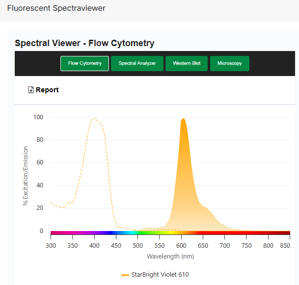
| 最大激发/发射波长 | 荧光染料: StarBright Violet 610 最大激发波长: 403 nm 最大发射波长: 607 nm |
| 监管声明 | 仅供研究使用 |
| 保质期 | 自发货之日起12个月 |
| 专利信息 | 本产品受美国专利号10,150,841及相关美国和外国对应专利的保护。 |
储存信息
本产品以环境温度运输。
请于 +4°C 储存。请勿冷冻。
本产品应储存于未稀释状态。
应用
本产品已报道适用于以下应用。此信息源自我们实验室的内部测试、同行评审的文献或原始作者的私人通讯。详细信息请参阅参考文献。如需通用实验方案建议,请访问抗体实验方案页面。
| 应用名称 | 已验证 | 推荐最低稀释度 | 推荐最高稀释度 |
|---|---|---|---|
| 流式细胞术 | ✓ | 原液 | - |
若本品未针对某项特定技术进行测试,并不代表其不适用于该技术。推荐的参考工作稀释度仅供参考,建议用户在其体系中通过适当的阴性/阳性对照,自行滴定确定最佳使用浓度。
流式细胞术
建议使用 5 µl 工作稀释液标记 100 µl 体积中的 10⁶ 个细胞。最佳实验流程建议在上样前以 6,000 g 离心 5 分钟。
CD14 is strongly expressed on the surface of monocytes and macrophages but has also been shown to be expressed on the surface of non-myeloid cells (Jersmann 2005). CD14 functions as a pattern recognition receptor (Pugin et al. 1994, Dziarski et al. 1998) in innate immunity for a variety of ligands, in particular for the LPS (endotoxin) of Gram-negative bacteria.
Mouse anti human CD14 antibody, clone TÜK4 has been shown to block SDF-induced chemotaxis of U937 cells in a dose –dependent manner (Yang et al. 2003). Use of the anti-human CD14 antibody, Low Endotoxin format is recommended for this purpose.
储存信息
Store at +4°C.DO NOT FREEZE.
This product should be stored undiluted.
应用
| Application Name | Verified | Min Dilution | Max Dilution |
|---|---|---|---|
| Flow Cytometry | ✓ | Neat |
Flow Cytometry
Use 5μl of the suggested working dilution to label 106 cells in 100μl. Best practices suggest a 5 minutes centrifugation at 6,000g prior to sample application.
风险提示:丁香通仅作为第三方平台,为商家信息发布提供平台空间。用户咨询产品时请注意保护个人信息及财产安全,合理判断,谨慎选购商品,商家和用户对交易行为负责。对于医疗器械类产品,请先查证核实企业经营资质和医疗器械产品注册证情况。
 文献和实验
文献和实验References for CD14 antibody
- Jacobsen, C.N. et al. (1993) Reactivities of 20 anti-human monoclonal antibodies with leucocytes from ten different animal species.
Vet Immunol Immunopathol. 39 (4): 461-6. - Gupta, V.K. et al. (1996) Identification of the sheep homologue of the monocyte cell surface molecule--CD14.
Vet Immunol Immunopathol. 51 (1-2): 89-99. - Sopp, P. & Howard, C.J. (1997) Cross-reactivity of monoclonal antibodies to defined human leucocyte differentiation antigens with bovine cells.
Vet Immunol Immunopathol. 56 (1-2): 11-25. - Werling, D. et al. (1998) Analysis of the phenotype and phagocytic activity of monocytes/macrophages from cattle infected with the bovine leukaemia virus.
Vet Immunol Immunopathol. 62 (3): 185-95. - Weiss, D.J. (2001) Evaluation of proliferative disorders in canine bone marrow by use of flow cytometric scatter plots and monoclonal antibodies.
Vet Pathol. 38: 512-8. - Bryan, S.A. et al. (2002) Responses of leukocytes to chemokines in whole blood and their antagonism by novel CC-chemokine receptor 3 antagonists.
Am J Respir Crit Care Med. 165: 1602-9. - Yang, H. et al. (2003) Antibody to CD14 like CXCR4-specific antibody 12G5 could inhibit CXCR4-dependent chemotaxis and HIV Env-mediated cell fusion.
Immunol Lett. 88 (1): 27-30. - Schenk, M. et al. (2005) Macrophages expressing triggering receptor expressed on myeloid cells-1 are underrepresented in the human intestine.
J Immunol. 174 (1): 517-24.
Further Reading
- Bazil, V. et al. (1986) Biochemical characterization of a soluble form of the 53-kDa monocyte surface antigen.
Eur J Immunol. 16:1583-9. - Simmons, D. L. et al. (1989) Monocyte antigen CD14 is a phospholipid anchored membrane protein.
Blood. 73:284-9. - Pugin, J. et al. (1994) CD14 is a pattern recognition receptor.
Immunity.1:509-16. - Dziarski, R. et al. (1998) Binding of bacterial peptidoglycan to CD14.
J Biol Chem. 273:8680-90. - Jersmann, H.P. (2005) Time to abandon dogma: CD14 is expressed by non-myeloid lineage cells.
Immunol Cell Biol. 83:462-7. - Piriou-Guzylack, L. (2008) Membrane markers of the immune cells in swine: an update.
Vet Res. 39: 54.
(共享) 高免血清制备的方案!! (共享) 免疫组化操作流程 (共享) 实验室分子生物学免疫学等常用试剂配方 (共享)92-99四军大免疫博士入学试题汇总! (共享)anti-CXCR1, anti-CXCR2, anti-CCR7 小鼠抗人单克隆抗体剩余 (共享)DNA疫苗技术 (共享)Eight Immunology flashs (共享)ELISA处理软件(1) (共享)elisa的通用规则 (共享)Elisa分析软件 (共享)ELISA数据处理软件(2) (共享
11c和CD123并不是DC特异的标志物。两类细胞均HLA-DR高表达,单核细胞、淋巴细胞和NK细胞的系列标记物弱表达。因此使用单一荧光标记的LIN cocktail(LIN1)抗体将DC细胞区分出来。LIN1抗体包括CD3、CD14、CD16、CD19、CD20和CD56。 另外,本实验还可以区分嗜碱粒细胞。嗜碱粒细胞的表型是:LIN1-CD123+HLA-DR-。使用HLA-DR可以区分嗜碱粒细胞和CD123+ DC细胞。 细胞来源: 样本使用EDTA、ACD或肝素钠抗凝全血,或分离的外周
恶性肿瘤病人外周血免疫指标的流式细胞仪检测及免疫治疗前后免疫功能变化的研究
,其中年龄和性别构成比与病人组差异无显著性(P>0.05)。 1.2 材料与仪器 小鼠抗人单克隆抗体CD4/CD8/CD3、CD19、CD20、CD3/CD16+56、CD3/HLA-DR、CD3/CD25及同型对照、红细胞裂解液(Optilyse C)均为法国Immunotech公司出品。流式细胞仪(Epics XL·MCL)是美国BECKMAN COULTER公司生产。 1.3 研究方法 使用直接免疫荧光标记全血溶血法,流式细胞仪测定肿瘤病人外周血淋巴细胞表面抗原表达。
 技术资料
技术资料暂无技术资料 索取技术资料

