

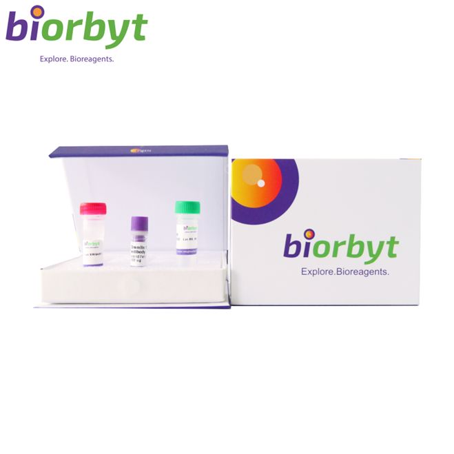
- ¥2808 - 7956
- Biorbyt

- orb1239348
- 英国
- 2026年07月29日
- ELISA, IF, IHC-P, KO/KD Validated, WB
- Rabbit
- Human, Mouse, Rat

企业认证
相关产品推荐更多 >
万千商家帮你免费找货
0 人在求购买到急需产品
相关阅读
香烟烟雾如何加速声带纤维化?CD44-透明质酸轴是关键通路Takara 秋促来袭,提取+反转录+定量套装仅 1200 元五大应用案例:Mustang Q 膜层析应用全解析1 个小工具,一次性搞定流程图、质粒图谱和信号通路图- 详细信息
- 技术资料
- 抗体名:
TNFRSF10B Antibody 抗体
- 抗体英文名:
TNFRSF10B Antibody
- 靶点:
TNFRSF10B
- 浓度:
1 mg/ml
- 应用范围:
ELISA, IF, IHC-P, KO/KD Validated, WB
- 宿主:
Rabbit
- 适应物种:
Human, Mouse, Rat
- 保质期:
12 months from date of receipt.
- 抗原来源:
详询
- 目录编号:
orb1239348
- 级别:
科研
- 库存:
99
- 供应商:
Biorbyt
- 标记物:
Unconjugated
- 克隆性:
Polyclonal
- 保存条件:
详询
- 形态:
Liquid
- 亚型:
IgG
- 免疫原:
详询
- 规格:
0.02 mg/0.1 mg
| 规格: | 0.02 mg | 产品价格: | ¥2808.0 |
|---|---|---|---|
| 规格: | 0.1 mg | 产品价格: | ¥7956.0 |
类别: Antibodies
产品子类型: Primary Antibody
描述: TNFRSF10B Antibody
别名: DR5 Antibody: DR5, CD262, KILLER, TRICK2, TRICKB, ZTNFR9, TRAILR2, TRICK2A, TRICK2B, TRAIL-R2, KILLER/DR5, DR5, UNQ160/PRO186, Tumor necrosis factor receptor superfamily member 10B, Death receptor 5, TRAIL receptor 2
应用说明: WB: 0.5-2 μg/mL; IHC-P: 5 μg/mL; IF: 5-10 μg/mL.Antibody validated: Western Blot in human, mouse and rat samples; Immunohistochemistry in mouse samples; Immunocytochemistry in human samples and Immunofluorescence in human, mouse and rat samples. All other applications and species not yet tested.
免疫原: DR5 antibody was raised against a peptide corresponding to 20 amino acids near the carboxy terminus of human DR5 precursor.The immunogen is located within the last 50 amino acids of DR5.
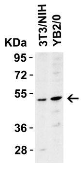
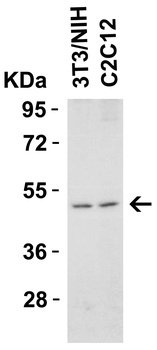
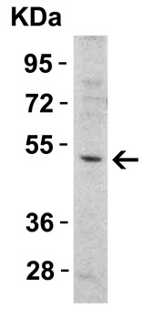
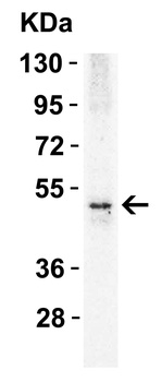
分子量: Predicted: 48kD and 45kDObserved: 48kD and 45kD
防腐剂: DR5 Antibody is supplied in PBS containing 0.02% sodium azide.
纯化方法: DR5 Antibody is affinity chromatography purified via peptide column.
特异性: Antibody has no cross reaction to DR4.
研究领域: Cell Biology
UniProt ID: O14763
NCBI 参考序列: AF012535
保存条件: Maintain refrigerated at 2-8°C for up to 2 weeks. For long term storage store at -20°C in small aliquots to prevent freeze-thaw cycles.
备注: For research use only.
产品子类型: Primary Antibody
描述: TNFRSF10B Antibody
别名: DR5 Antibody: DR5, CD262, KILLER, TRICK2, TRICKB, ZTNFR9, TRAILR2, TRICK2A, TRICK2B, TRAIL-R2, KILLER/DR5, DR5, UNQ160/PRO186, Tumor necrosis factor receptor superfamily member 10B, Death receptor 5, TRAIL receptor 2
应用说明: WB: 0.5-2 μg/mL; IHC-P: 5 μg/mL; IF: 5-10 μg/mL.Antibody validated: Western Blot in human, mouse and rat samples; Immunohistochemistry in mouse samples; Immunocytochemistry in human samples and Immunofluorescence in human, mouse and rat samples. All other applications and species not yet tested.
免疫原: DR5 antibody was raised against a peptide corresponding to 20 amino acids near the carboxy terminus of human DR5 precursor.The immunogen is located within the last 50 amino acids of DR5.
分子量: Predicted: 48kD and 45kDObserved: 48kD and 45kD
防腐剂: DR5 Antibody is supplied in PBS containing 0.02% sodium azide.
纯化方法: DR5 Antibody is affinity chromatography purified via peptide column.
特异性: Antibody has no cross reaction to DR4.
研究领域: Cell Biology
UniProt ID: O14763
NCBI 参考序列: AF012535
保存条件: Maintain refrigerated at 2-8°C for up to 2 weeks. For long term storage store at -20°C in small aliquots to prevent freeze-thaw cycles.
备注: For research use only.
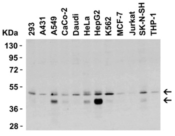
Western Blot Validation in Human Cell Lines. Loading: 15 µg of lysates per lane. Antibodies: DR5 orb1239348, (0.5 µg/mL), 1h incubation at RT in 5% NFDM/TBST. Secondary: Goat anti-rabbit IgG HRP conjugate at 1:10000 dilution.
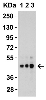
Western Blot Validation in Human HepG2 Cells. Loading: 15 µg of lysates per lane. Antibodies: DR5 orb1239348, 1h incubation at RT in 5% NFDM/TBST. Secondary: Goat anti-rabbit IgG HRP conjugate at 1:10000 dilution. Lane 1: 1 µg/mL, Lane 2: 2 µg/mL, Lane 3: 4 µg/mL.
Western Blot Validation in Mouse and Rat Cell Lines. Loading: 15 µg of lysates per lane. Antibodies: DR5 orb1239348, (2 µg/mL), 1h incubation at RT in 5% NFDM/TBST. Secondary: Goat anti-rabbit IgG HRP.
Western Blot Validation in Mouse Cell Lines. Loading: 15 µg of lysates per lane. Antibodies: DR5 orb1239348, (1 µg/mL), 1h incubation at RT in 5% NFDM/TBST. Secondary: Goat anti-rabbit IgG HRP conjugate at 1:10000 dilution.
Western Blot Validation in Mouse Heart. Loading: 15 µg of lysatesper lane. Antibodies: DR5 orb1239348, (1 µg/mL), 1h incubation at RT in 5% NFDM/TBST. Secondary: Goat anti-rabbit IgG HRP conjugate at 1:10000 dilution.
Western Blot Validation in Rat Skeletal Muscle. Loading: 15 µg of lysate per lane. Antibodies: DR5 orb1239348, (1 µg/mL), 1h incubation at RT in 5% NFDM/TBST. Secondary: Goat anti-rabbit IgG HRP conjugate at 1:10000 dilution.
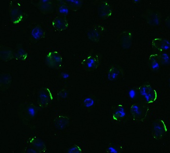
Immunofluorescence Validation of DR5 in Human HepG2 Cells. Immunofluorescent analysis of 4% Fixative-fixed human HepG2 cells labeling DR5 with orb1239348 at 5 µg/mL, followed by goat anti-rabbit IgG secondary antibody at 1/500 dilution (green) and DAPI (blue).
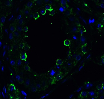
Immunofluorescence Validation of DR5 in Human Testis. Immunofluorescent analysis of 4% Fixative-fixed human testis tissue labeling DR5 with orb1239348 at 10 µg/mL, followed by goat anti-rabbit IgG secondary antibody at 1/500 dilution (green) and DAPI (blue).
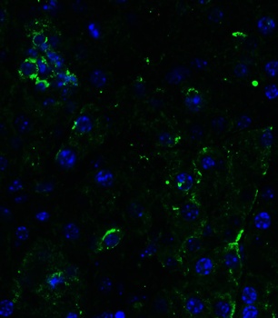
Immunofluorescence Validation of DR5 in Mouse Pancreas. Immunofluorescent analysis of 4% Fixative-fixed mouse pancreas tissue labeling DR5 with orb1239348 at 10 µg/mL, followed by goat anti-rabbit IgG secondary antibody at 1/500 dilution (green) and DAPI (blue).
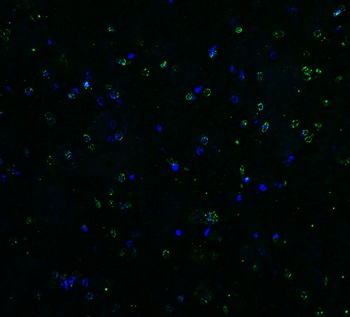
Immunofluorescence Validation of DR5 in Rat Brain. Immunofluorescent analysis of 4% Fixative-fixed rat brain tissue labeling DR5 with orb1239348 at 5 µg/mL, followed by goat anti-rabbit IgG secondary antibody at 1/500 dilution (green) and DAPI (blue).
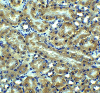
Immunohistochemistry Validation of DR5 in Mouse kidney tissue. Immunohistochemical analysis of paraffin-embedded mouse kidney tissue using anti-DR5 antibody (orb1239348) at 5 µg/ml. Tissue was fixed with formaldehyde and blocked with 10% serum for 1 h at RT; antigen retrieval was by heat mediation with a citrate buffer (pH6). Samples were incubated with primary antibody overnight at 4°C. A goat anti-rabbit IgG H&L (HRP) at 1/250 was used as secondary. Counter stained with Hematoxylin.
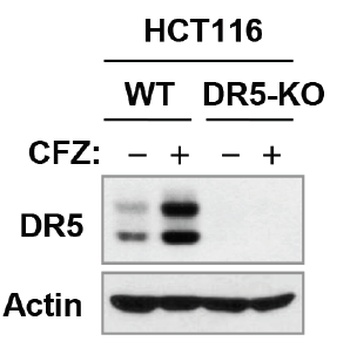
KO Validation of DR5 in HCT116 Cells (Han et al., 2015). Anti-cancer drug, Carfilzomib (CFZ), induced up-regulation of DR5 and the expression of DR5 was not detected in DR5-KO HCT 116 cell line with anti-DR5 antibodies (orb1239348).
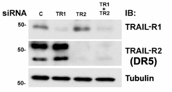
KD Validation of DR5 in MB231 Cells (Rahman et al., 2009). Western blot analysis with anti-DR5 antibodies was performed for DR5 in MB231 cells transfected with control siRNA or DR5 siRNA. DR5 expression was disrupted after DR5 siRNA knockdown.
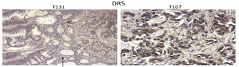
Immunohistochemistry Validation of BIM in Human Colon Tumors (Devetzi et al., 2016). Protein analysis for DR5 by immunohistochemistry with anti-DR5 antibodies in human colon tumors. Strong immunoreactivity is shown for DR5 in T167 patient with colorectal cancer.
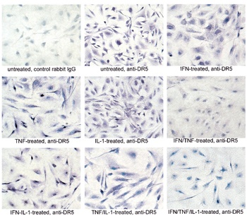
Regulated Expression Validation of DR5 in Thyroid Epithelial Cells (Bretz et al., 2002). Immunostaining with anti-DR5 antibodies shows high levels of DR5 expression in untreated cells and cells treated with each of the three cytokines alone or TNFalpha combined with IL-1b. In contrast, treatment with both IFNg and TNFalpha or all three cytokines greatly reduces DR5 staining. The reduction in staining appears most significant in cytoplasmic regions while some staining is maintained in or around the nucleus.
风险提示:丁香通仅作为第三方平台,为商家信息发布提供平台空间。用户咨询产品时请注意保护个人信息及财产安全,合理判断,谨慎选购商品,商家和用户对交易行为负责。对于医疗器械类产品,请先查证核实企业经营资质和医疗器械产品注册证情况。
 技术资料
技术资料暂无技术资料 索取技术资料
TNFRSF10B Antibody 抗体,orb1239348,Biorbyt
¥2808 - 7956

