产品描述:Mouse monoclonal to Kir2 (RPE). 1. Ion channels are integral membrane proteins that help establish and control the small voltage gradient across the plasma membrane of living cells by allowing the flow of ions down their electrochemical gradient. They are present in the membranes that surround all biological cells because their main function is to regulate the flow of ions across this membrane. Whereas some ion channels permit the passage of ions based on charge, others conduct based on a ionic species, such as sodium or potassium. Furthermore, in some ion channels, the passage is governed by a gate which is controlled by chemical or electrical signals, temperature, or mechanical forces. There are a few main classifications of gated ion channels. There are voltage- gated ion channels, ligand- gated, other gating systems and finally those that are classified differently, having more exotic characteristics. The first are voltage- gated ion channels which open and close in response to membrane potential. These are then separated into sodium, calcium, potassium, proton, transient receptor, and cyclic nucleotide-gated channels; each of which is responsible for a unique role. Ligand-gated ion channels are also known as ionotropic receptors, and they open in response to specific ligand molecules binding to the extracellular domain of the receptor protein. The other gated classifications include activation and inactivation by second messengers, inward-rectifier potassium channels, calcium-activated potassium channels, two-pore-domain potassium channels, light-gated channels, mechano-sensitive ion channels and cyclic nucleotide-gated channels. Finally, the other classifications are based on less normal characteristics such as two-pore channels, and transient receptor potential channels. The Kir2. 1 inward-rectifier potassium ion channel is encoded by the KCNJ2 gene. A defect in this gene is associated with Andersen-Tawil syndrome....
别名:Kir2.1, KCNJ2, Inward rectifier potassium channel 2, Inward rectifier K(+) channel Kir2.1, Potassium channel inwardly rectifying subfamily J member 2, IRK1, HHBIRK1, HHIRK1, HIRK 1, LQT7, SQT3
免疫原:Fusion protein amino acids 41-64 and 189-428 of mouse Kir2.1
克隆性:S112
分子量:45kDa
应用稀释比例:WB (1:1000), IHC (1:1000), ICC/IF (1:100)
应用注释:1 µg/ml of SMC-310 was sufficient for detection of Kir2.1 in 10 µg of rat brain lysate by colorimetric immunoblot analysis using Goat anti-mouse IgG:HRP as the secondary antibody.
防腐剂:95.46mM Phosphate, 2.48mM MES and 2mM EDTA
纯化:Protein G Purified
保存说明:Conjugated antibodies should be stored according to the product label
NCBI:NP_032451
Entrez:16518
UniProt ID:P35561
Note:For research use only.
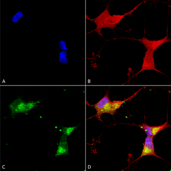
Immunocytochemistry/Immunofluorescence analysis using Mouse Anti-Kir2.1 Monoclonal Antibody, Clone S112. Tissue: Neuroblastoma cells (SH-SY5Y). Species: Human. Fixation: 4% PFA for 15 min. Primary Antibody: Mouse Anti-Kir2.1 Monoclonal Antibody at 1:50 for overnight at 4°C with slow rocking. Secondary Antibody: AlexaFluor 488 at 1:1000 for 1 hour at RT. Counterstain: Phalloidin-iFluor 647 (red) F-Actin stain; Hoechst (blue) nuclear stain at 1:800, 1.6mM for 20 min at RT. (A) Hoechst (blue) nuclear stain. (B) Phalloidin-iFluor 647 (red) F-Actin stain. (C) Kir2.1 Antibody (D) Composite.
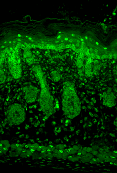
Immunohistochemistry analysis using Mouse Anti-Kir2.1 Potassium Channel Monoclonal Antibody, Clone S112. Tissue: backskin. Species: Mouse. Fixation: Bouin's Fixative and paraffin-embedded. Primary Antibody: Mouse Anti-Kir2.1 Potassium Channel Monoclonal Antibody at 1:100 for 1 hour at RT. Secondary Antibody: FITC Goat Anti-Mouse (green) at 1:50 for 1 hour at RT. Localization: Nuclear expression in the epidermis and hair follicles.
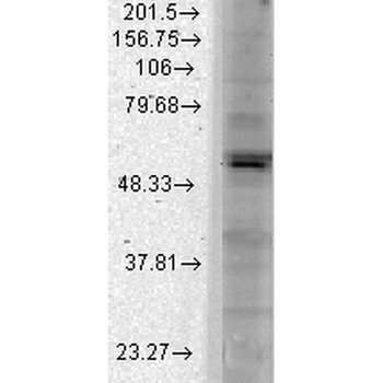
Western Blot analysis of Monkey COS transient cell lysate showing detection of Kir2.1 Potassium Channel protein using Mouse Anti-Kir2.1 Potassium Channel Monoclonal Antibody, Clone S112. Load: 15 μg. Block: 1.5% BSA for 30 minutes at RT. Primary Antibody: Mouse Anti-Kir2.1 Potassium Channel Monoclonal Antibody at 1:1000 for 2 hours at RT. Secondary Antibody: Sheep Anti-Mouse IgG: HRP for 1 hour at RT.
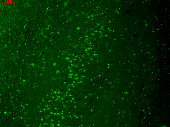
Immunohistochemistry analysis using Mouse Anti-Kir2.1 Potassium Channel Monoclonal Antibody, Clone S112. Tissue: hippocampus. Species: Human. Fixation: Bouin's Fixative and paraffin-embedded. Primary Antibody: Mouse Anti-Kir2.1 Potassium Channel Monoclonal Antibody at 1:1000 for 1 hour at RT. Secondary Antibody: FITC Goat Anti-Mouse (green) at 1:50 for 1 hour at RT.


 文献和实验
文献和实验 技术资料
技术资料
