


- ¥12000
- 苏州艾洛蒙生物
- ARD0369
- 苏州
- 2025年12月18日
- Human

企业认证
相关产品推荐更多 >

万千商家帮你免费找货
0 人在求购买到急需产品
- 详细信息
- 技术资料
- 抗体名:
菲达斯妥单抗
- 抗体英文名:
Fidasimtamab Biosimilar
- 靶点:
ERBB2 (epidermal growth factor receptor 2, receptor tyrosine-protein kinase erbB-2, EGFR2, HER2, HER-2, p185c-erbB2, NEU,CD340) [Homo sapiens],PDCD1 (programmed cell death 1, PD1, PD-1, CD279) [Homo sapiens] bispecific
- 适应物种:
Human
- 保质期:
12 months
- 目录编号:
CAS:2377419-89-9
- 级别:
Research Grade
- 供应商:
苏州艾洛蒙
- 标记物:
无
- 克隆性:
Monoclonal
- 保存条件:
Store at -20°C for 12 months (Avoid repeated freezing and thawing)
- 形态:
Liquid
- 亚型:
Human IgG1, κ
- 规格:
1mg
Fidasimtamab (菲达斯妥单抗;IBI-315; BH2950)为一重组人IgG1双特异性抗体,能同时针对HER2及PD-1进行靶向、结合与抑制,进而影响其下游信号通路。此外,该物质通过连接表达PD-1的T细胞与表达HER2的肿瘤细胞,展现其潜在的免疫抑制及抗肿瘤活性。Fidasimtamab (IBI-315; BH2950), a recombinant human IgG1 bispecific antibody, specifically targets, binds to, and inhibits HER2 and PD-1 along with their downstream signaling pathways. Additionally, it bridges PD-1 expressing T cells with HER2 expressing tumor cells, demonstrating potential immunosuppressive and antitumor activities。
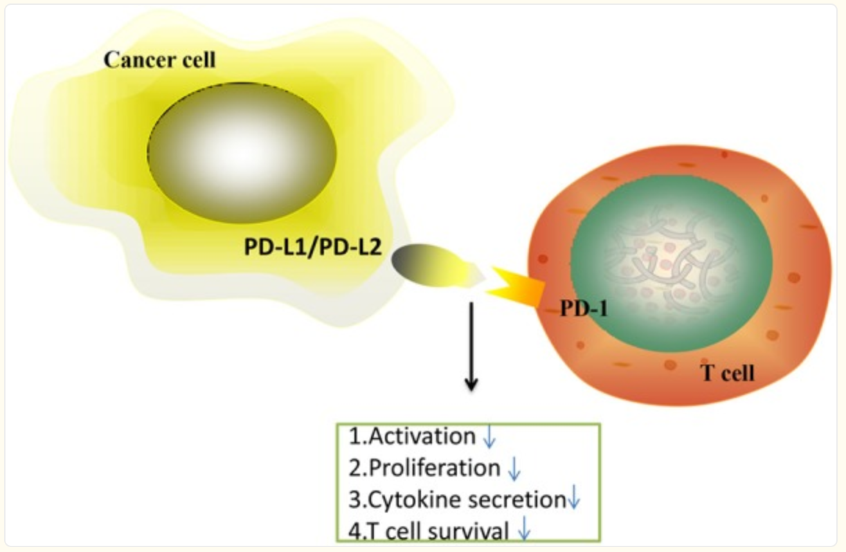
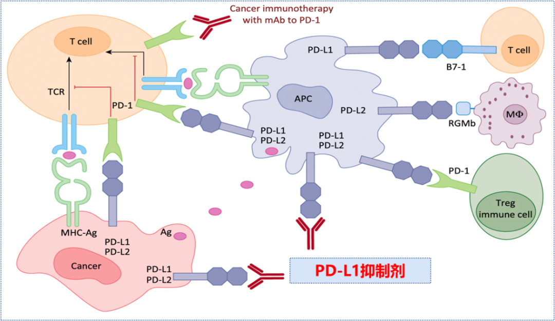
在过去的几年里,癌症免疫疗法一直伴随着有希望的结果。程序性细胞死亡蛋白1(Programmed Cell Death Protein 1,PD-1)通过调节T细胞活性、激活抗原特异性T细胞凋亡和抑制调节性T细胞凋亡,在抑制免疫应答和促进自身免疫耐受中发挥重要作用。程序性细胞死亡配体1(Programmed Cell Death Ligand 1,PD-L1)是一种跨膜蛋白,被认为是免疫应答的共抑制因子,它能与PD-1联合收割机结合,减少PD-1阳性细胞的增殖,抑制其细胞因子分泌,诱导细胞凋亡。PD-L1还在各种恶性肿瘤中发挥重要作用,它可以减弱宿主对肿瘤细胞的免疫反应。基于这些观点,PD-1/PD-L1轴负责癌症免疫逃逸,并对癌症治疗产生巨大影响。本文就PD-1和PD-L1在肿瘤中的作用作一综述,以期为肿瘤的治疗提供参考。
PD-1(programmed cell death protein 1)是程序性死亡受体1,是活化的T细胞表达的免疫检查点受体,是一种重要的免疫抑制分子。PD-1通过向下调节免疫系统对人体细胞的反应,以及通过抑制T细胞炎症活动来调节免疫系统并促进自身耐受。这可以预防自身免疫疾病,但它也可以防止免疫系统杀死癌细胞。 PD-L1是PD-1的配体,PD-L1与免疫系统的抑制有关,可以传导抑制性的信号。肿瘤细胞也可以表达PD-L1,PD-1和PD-L1一旦结合便会向T细胞传递负向调控信号,导致T细胞无法识别癌细胞,肿瘤细胞从而实现“免疫逃逸”。 PD-1可表达在T细胞、B细胞等免疫细胞表面。不过在T细胞未活化时,几乎是不表达PD-1的,仅在T细胞活化之后,PD-1才表达在T细胞表面。 PD-L1除了会表达在肿瘤表面,参与免疫逃逸,还会在IFN-γ刺激下,表达在抗原提呈细胞(DC细胞、巨噬细胞等),以及血管内皮细胞的表面。
Cancer immunotherapy has been accompanied by promising results over the past few years. Programmed Cell Death Protein 1 (PD-1) plays a vital role in inhibiting immune responses and promoting self-tolerance through modulating the activity of T-cells, activating apoptosis of antigen-specific T cells and inhibiting apoptosis of regulatory T cells. Programmed Cell Death Ligand 1 (PD-L1) is a trans-membrane protein that is considered to be a co-inhibitory factor of the immune response, it can combine with PD-1 to reduce the proliferation of PD-1 positive cells, inhibit their cytokine secretion and induce apoptosis. PD-L1 also plays an important role in various malignancies where it can attenuate the host immune response to tumor cells. Based on these perspectives, PD-1/PD-L1 axis is responsible for cancer immune escape and makes a huge effect on cancer therapy. This review is aimed to summarize the role of PD-1 and PD-L1 in cancer, looking forward to improve the therapy of cancer.
人表皮生长因子受体2(human epidermal growth factor 2,HER2)基因,即酪氨酸激酶受体2(Erb-B2 receptor tyrosine kinase 2,ERBB2)基因,位于人17号染色体q12-21,编码跨膜酪氨酸激酶受体蛋白。HER2基因扩增及蛋白的过度表达会传递信号,刺激细胞增值。
HER2也称为c-erB2,包括三个结构域:胞外结合域(the extracellular domain,ECD)、跨膜结构域(a transmembrane domain,TM)和胞内结构域(an intrcellular domain,ICD)。胞外区可分4个亚结构域(I-IV),I、III亚结构域为配体的结合位点,II、IV亚结构域存在丰富的半胱氨酸,可以形成同源或异源二聚体。跨膜区为α螺旋结构。胞内区包含了多个重要的环状结构,构成酪氨酸激酶的活性位点。人类该基因定位于染色体17q21,属于原癌基因。其编码产物HER2蛋白为185kD的跨膜精蛋白,简称p185,由1255个氨基酸组成,720—987位属于酪氨酸激酶区。HER2蛋白是具有酪氨酸蛋白激酶活性的跨膜蛋白,属于EGFR家族成员之一。蛋白由胞外的配体结合区、单链跨膜区及胞内的蛋白酪氨酸激酶区三部分组成,由于尚未发现能与其直接结合的配体,HER2蛋白主要通过与家族中其他成员包括EGFR(HERl/erbBI)、HER3/erbB3、HER4/erbB4形成异二聚体而与各自的配体结合。HER2蛋白常为异二聚体,且活性常强于其它异二聚体。当与配体结合后,主要通过引起受体二聚化及胞浆内酪氨酸激酶区的自身磷酸化,激活酪氨酸激酶的活性。HER2蛋白介导的信号转导途径主要有Ras/Raf/分裂素活化蛋白激酶(MAPK)途径,磷脂酰肌醇3羟基激酶(PI3K)/Akt途径,信号转导及转录激活(STAT)途径和PLC通路等。
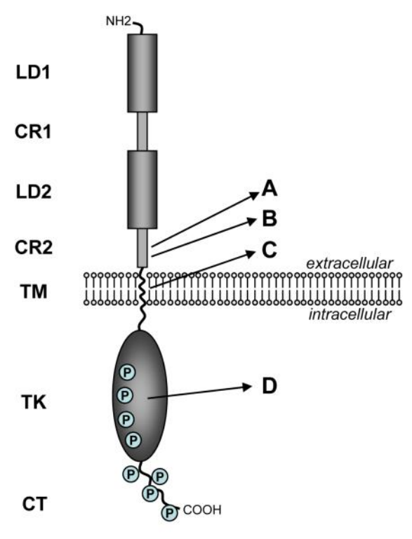
Structure of the HER2 and Neu proteins. The domain structure is shown on the left consisting of two ligand binding regions (LD1 & LD2), two cysteine-rich regions (CR1 & CR2), a short transmembrane domain (TM), a catalytic tyrosine kinase domain (TK), and a carboxy terminal tail (CT). Numerous sites of tyrosine phosphorylation wiithin the TK and CT domains are indicated by circled P.The letters on the right point to specific areas that are altered or mutated in certain naturally occuring or experimentally induced cancers discussed in the text. A) site of somatic mutations found in tumors arising in MMTV-neu mice. B) site of the 48bp deletion in the naturally occuring human ΔHER2 isoform. C) site of the mutation in the neuT oncogene initially discovered in a rat carcinogen induced tumor model and subsequently used in numerous in vitro and transgenic experimental models. D) site of mutations found in rare cases of human lung cancers.
风险提示:丁香通仅作为第三方平台,为商家信息发布提供平台空间。用户咨询产品时请注意保护个人信息及财产安全,合理判断,谨慎选购商品,商家和用户对交易行为负责。对于医疗器械类产品,请先查证核实企业经营资质和医疗器械产品注册证情况。
 技术资料
技术资料暂无技术资料 索取技术资料
