


- ¥1480
- EK-Bioscience

- CCC-Y4007
- 2026年05月11日

企业认证
相关产品推荐更多 >

万千商家帮你免费找货
0 人在求购买到急需产品
- 详细信息
- 文献和实验
- 技术资料
- 规格:
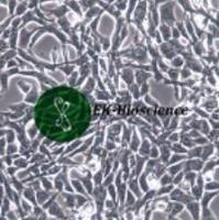
T25
MDCK/MDCK细胞系/MDCK细胞株/MDCK犬肾上皮细胞
Cell line name MDCK
Synonyms MDCK (NBL-2); MDCK(NBL-2); NBL-2; Madin-Darby Canine Kidney; Madin Darby Canine Kidney
Accession CVCL_0422
Resource Identification Initiative To cite this cell line use: MDCK (RRID:CVCL_0422)
Comments Group: Vaccine production cell line.
Part of: Naval Biosciences Laboratory (NBL) collection (transferred to ATCC in 1982).
Characteristics: Used for virus production for vaccine development, toxicology and industrial biotechnology research, and high-throughput screening (ATCC=CCL-34).
Virology: Not susceptible to infection by SARS coronavirus 2 (SARS-CoV-2) (COVID-19) (PubMed=33389257).
Omics: Transcriptome analysis by RNAseq.
Caution: There seem to be two distinct cell lines which were assigned NCBI_Iran catalog number C426.
Derived from site: In situ; Kidney; UBERON=UBERON_0002113.
Cell type: Epithelial cell of kidney; CL=CL_0002518.
Species of origin Canis lupus familiaris (Dog) (Canis familiaris) (NCBI Taxonomy: 9615)
Breed/subspecies: Cocker Spaniel; VBO=VBO_0200372.
Hierarchy Children:
Age at sampling Adult
Category Spontaneously immortalized cell line
PubMed=13901412; DOI=10.1126/science.138.3536.42
Green I.J.
Serial propagation of influenza B (Lee) virus in a transmissible line of canine kidney cells.
Science 138:42-43(1962)
PubMed=5918973; DOI=10.3181/00379727-122-31293
Gaush C.R., Hard W.L., Smith T.F.
Characterization of an established line of canine kidney cells (MDCK).
Proc. Soc. Exp. Biol. Med. 122:931-935(1966)
DOI=10.1007/BF02618370
Stulberg C.S., Coriell L.L., Kniazeff A.J., Shannon J.E.
The animal cell culture collection.
In Vitro 5:1-16(1970)
PubMed=222773; DOI=10.1083/jcb.81.3.635; PMCID=PMC2110404
Rindler M.J., Chuman L.M., Shaffer L., Saier M.H. Jr.
Retention of differentiated properties in an established dog kidney epithelial cell line (MDCK).
J. Cell Biol. 81:635-648(1979)
PubMed=6480288
Schroyens W., Bruyneel R., Tchao R., Leighton J., Dragonetti C.H., Mareel M.M.
Comparison of invasiveness and non-invasiveness of two epithelial cell lines in vitro.
Invasion Metastasis 4:160-170(1984)
PubMed=12667817; DOI=10.1016/S0042-6822(02)00064-8
Romanova J., Katinger D., Ferko B., Voglauer R., Mochalova L., Bovin N., Lim W., Katinger H., Egorov A.
Distinct host range of influenza H3N2 virus isolates in Vero and MDCK cells is determined by cell specific glycosylation pattern.
Virology 307:90-97(2003)
PubMed=19941903; DOI=10.1016/j.jviromet.2009.11.022
Karger A., Bettin B., Lenk M., Mettenleiter T.C.
Rapid characterisation of cell cultures by matrix-assisted laser desorption/ionisation mass spectrometric typing.
J. Virol. Methods 164:116-121(2010)
PubMed=21819694; PMCID=PMC3123757
Omeir R.L., Teferedegne B., Foseh G.S., Beren J.J., Snoy P.J., Brinster L.R., Cook J.L., Peden K., Lewis A.M. Jr.
Heterogeneity of the tumorigenic phenotype expressed by Madin-Darby canine kidney cells.
Comp. Med. 61:243-250(2011)
PubMed=21982418; DOI=10.1186/1471-2121-12-43; PMCID=PMC3209442
Dukes J.D., Whitley P., Chalmers A.D.
The MDCK variety pack: choosing the right strain.
BMC Cell Biol. 12:43.1-43.4(2011)
PubMed=24058646; DOI=10.1371/journal.pone.0075014; PMCID=PMC3772841
Lugovtsev V.Y., Melnyk D., Weir J.P.
Heterogeneity of the MDCK cell line and its applicability for influenza virus research.
PLoS ONE 8:E75014-E75014(2013)
PubMed=25903999; DOI=10.1002/biot.201400388
Genzel Y.
Designing cell lines for viral vaccine production: where do we stand?
Biotechnol. J. 10:728-740(2015)
PubMed=32854295; DOI=10.3390/ijms21176111; PMCID=PMC7504089
Rodrigues A.F., Fernandes P., Laske T., Castro R., Alves P.M., Genzel Y., Coroadinha A.S.
Cell bank origin of MDCK parental cells shapes adaptation to serum-free suspension culture and canine adenoviral vector production.
Int. J. Mol. Sci. 21:6111.1-6111.27(2020)
PubMed=33122286; DOI=10.1681/ASN.2020010009; PMCID=PMC7894662
Khundmiri S.J., Chen L.-H., Lederer E.D., Yang C.-R., Knepper M.A.
Transcriptomes of major proximal tubule cell culture models.
J. Am. Soc. Nephrol. 32:86-97(2021)
PubMed=33389257; DOI=10.1007/s10096-020-04106-0; PMCID=PMC7778494
Wurtz N., Penant G., Jardot P., Duclos N., La Scola B.
Culture of SARS-CoV-2 in a panel of laboratory cell lines, permissivity, and differences in growth profile.
Eur. J. Clin. Microbiol. Infect. Dis. 40:477-484(2021)
风险提示:丁香通仅作为第三方平台,为商家信息发布提供平台空间。用户咨询产品时请注意保护个人信息及财产安全,合理判断,谨慎选购商品,商家和用户对交易行为负责。对于医疗器械类产品,请先查证核实企业经营资质和医疗器械产品注册证情况。
 文献和实验
文献和实验*发表【中文论文】请标注:由上海酶研生物科技有限公司提供;
*发表【英文论文】请标注:From Shanghai EK-Bioscience Biotechnology Co., Ltd.
一、目的MDCK细胞培养是分离流感病毒及相关研究实验的基本技术。疾控中心的所有技术人员,必须按照本文件相关的操作规程进行操作。二、适用范围适用于疾控中心所有技术人员 。三、程序(一)生物安全要求实验室生物安全级别:BSL-1所有操作必须在BSL-1实验室的生物安全柜里进行。(二)材料1. 生长成片的MDCK细胞2. 无菌的T25细胞培养瓶3. D-MEM培养液(含有L-谷氨酰胺)4. 青、链霉素母液(10000 U/mL青霉素G;10000µg/mL硫酸链霉素),分装后保存于-20
一、目的MDCK细胞培养是分离流感病毒及相关研究实验的基本技术。疾控中心的所有技术人员,必须按照本文件相关的操作规程进行操作。二、适用范围适用于疾控中心所有技术人员 。三、程序(一)生物安全要求实验室生物安全级别:BSL-1所有操作必须在BSL-1实验室的生物安全柜里进行。(二)材料1. 生长成片的MDCK细胞2. 无菌的T25细胞培养瓶3. D-MEM培养液(含有L-谷氨酰胺)4. 青、链霉素母液(10000 U/mL青霉素G;10000µg/mL硫酸链霉素),分装后保存于-20
一、目的流感病毒的MDCK细胞分离方法是流感实验的关键技术,用于流感病毒的分离、培养。本SOP是为确保MDCK细胞分离流感病毒的操作准确、规范和可靠而制定。二、范围适用于实验室所有技术人员进行流感病毒的MDCK细胞分离。三、程序(一)生物安全要求H5,H7亚型高致病性禽流感病毒,H2N2亚型流感病毒的MDCK细胞分离实验室生物安全级别:BSL-3。实验操作人员需进行BSL-3防护,具体详见“生物安全个人防护SOP”。H5,H7亚型高致病性禽流感病毒,H2N2亚型流感病毒的MDCK细胞分离操作
 技术资料
技术资料
