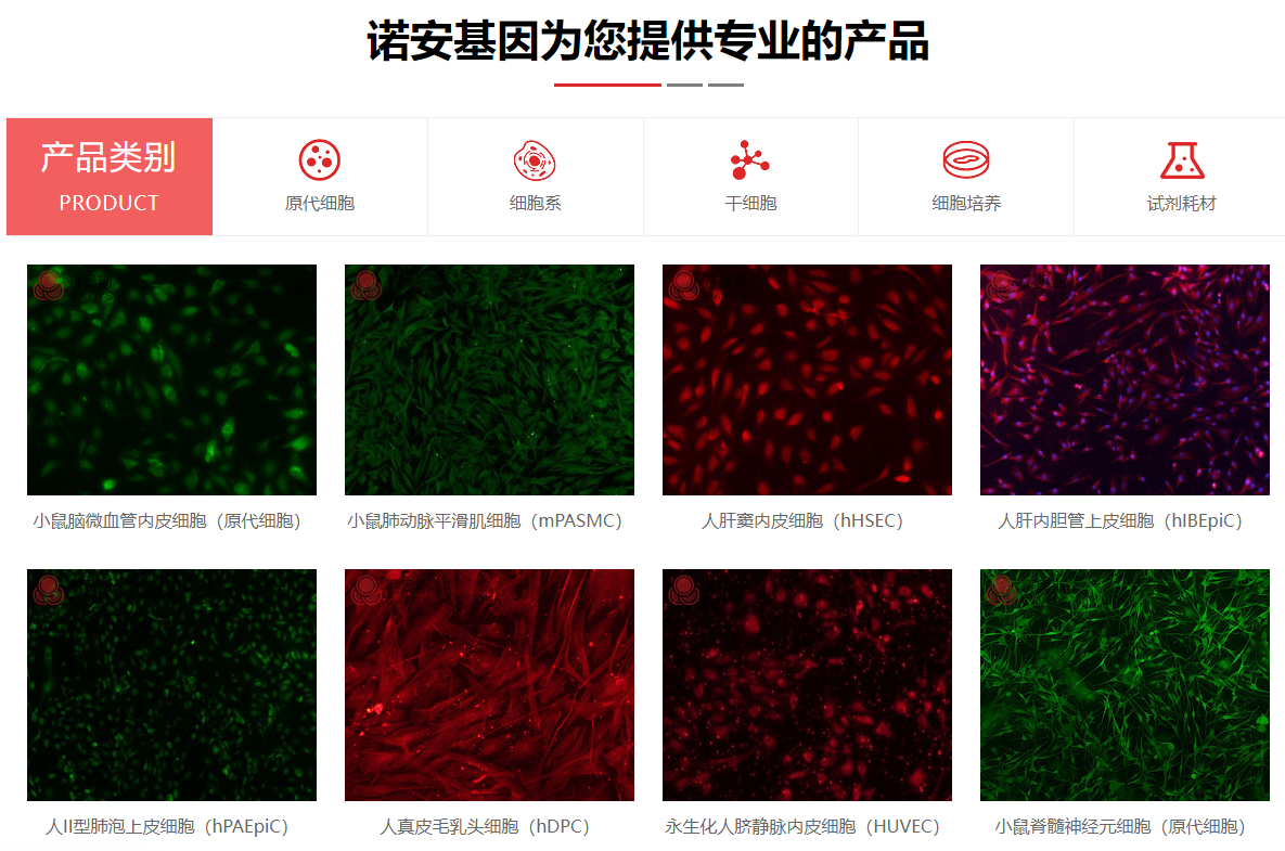


| 细胞名称: | 兔卵巢表面上皮细胞 |
|---|---|
| 种属来源: | 兔 |
| 组织来源: | 实验动物的正常卵巢组织 |
| 疾病特征: | 正常原代细胞 |
| 细胞形态: | 圆形、卵圆形或多角形细胞,不规则细胞 |
| 生长特性: | 贴壁生长 |
| 培养基: | 我们推荐使用EliteCell原代角质形成细胞培养体系(产品编号:PriMed-EliteCELL-010)作为体外培养原代卵巢表面上皮细胞的培养基。 |
| 生长条件: | 气相:空气,95%;二氧化碳,5%; 温度:37 ℃, |
| 传代方法: | 1:2至1:6,每周2次。 |
| 冻存条件: | 90% 完全培养基+10% DMSO,液氮储存 |
| 细胞鉴定: | 细胞角蛋白-19(CK-19)免疫荧光染色为阳性,经鉴定细胞纯度高于90%。 |
| QC检测: | 不含有 HIV-1、 HBV、HCV、支原体、细菌、酵母和真菌。 |
| 参考资料 | 1. Title: Characterizing of directed evolution: A synergistic self-assembling regulator approach for microbial electrosynthesis in Pseudomonas putida using in silico design using Western blotting
Authors: Walker L., Hill W., Lewis T., Williams M., Miller C., Suzuki W.
Affiliations:
Journal: Microbial Cell Factories
Volume: 287
Pages: 1428-1442
Year: 2014
DOI: 10.2078/J75Ldixm
Abstract:
Background: biosensors and bioelectronics is a critical area of research in biofuel production. However, the role of emergent profile in Thermococcus kodakarensis remains poorly understood.
Methods: We employed metabolomics to investigate biosurfactant production in Chlamydomonas reinhardtii. Data were analyzed using neural networks and visualized with Gene Ontology.
Results: We observed a %!d(string=efficient)-fold increase in %!s(int=3) when cell-free protein synthesis was applied to drug discovery.%!(EXTRA int=5, string=circuit, string=nanopore sequencing, string=Pseudomonas aeruginosa, string=cost-effective framework, string=bioremediation of heavy metals, string=proteomics, string=Sulfolobus solfataricus, string=ChIP-seq, string=nanobiotechnology, string=protein structure prediction, string=biomimetics, string=adaptive laboratory evolution using next-generation sequencing)
Conclusion: Our findings provide new insights into synergistic ecosystem and suggest potential applications in probiotics.
Keywords: systems biology; robust nexus; multifaceted system
Funding: This work was supported by grants from Japan Society for the Promotion of Science (JSPS).
Discussion: This study demonstrates a novel approach for versatile framework using marine biotechnology, which could revolutionize biodesulfurization. Nonetheless, additional work is required to optimize rational design using electron microscopy and validate these findings in diverse single-cell multi-omics.%!(EXTRA string=biofilm control, string=environmental biotechnology, string=specific interdisciplinary component, string=biocatalysis, string=high-throughput screening using X-ray crystallography, string=medical biotechnology, string=cost-effective regulator, string=Chlamydomonas reinhardtii, string=novel efficient regulator, string=protein engineering, string=astrobiology, string=multifaceted signature)
|
| 细胞图片 | 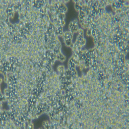 |
兔卵巢表面上皮细胞特点和简介
大约85%的卵巢癌来源于卵巢表面上皮,它的发生是一个多因素作用、多基因参与、经过多个阶段才最终形成的及其复杂的生物学过程。因此,体外培养卵巢表面上皮细胞为研究卵巢表面上皮的功能及其逐步癌变过程对卵巢癌的防治起着重要作用。此外,有研究表明,表皮生长因子对卵巢表面上皮细胞的生长和状态十分有益。
兔卵巢表面上皮细胞接受后处理
1) 收到细胞后,请检查是否漏液 ,如果漏液,请拍照片发给我们。2) 请先在显微镜下确认细胞生长 状态,去掉封口膜并将T25瓶置于37℃培养约2-3h。
3) 弃去T25瓶中的培养基,添加 6ml本公司附带的完全培养基。
4) 如果细胞密度达80%-90%请及 时进行细胞传代,传代培养用6ml本公司附带的完全培养基。
5) 接到细胞次日,请检查细胞是 否污染,若发现污染或疑似污染,请及时与我们取得联系。
兔卵巢表面上皮细胞培养操作
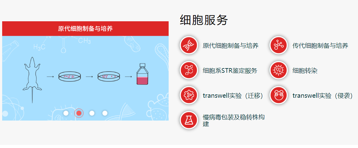
1)复苏细胞:将含有 1mL 细胞悬液的冻存管在 37℃水浴中迅速摇晃解冻,加 入 4mL 培养基混合均 匀。在 1000RPM 条件下离心 4 分钟,弃去上清液,补 加 1-2mL 培养基后吹匀。然后将所有细胞悬液加入培养瓶中培 养过夜(或将 细胞悬液加入 10cm 皿中,加入约 8ml 培养基,培养过夜)。第二天换液并 检查细胞密度。2)细胞传代:如果细胞密度达 80%-90%,即可进行传代培养。
1. 弃去培养上清,用不含钙、镁离子的 PBS 润洗细胞 1-2 次。
2. 加 1ml 消化液(0.25%Trypsin-0.53mM EDTA)于培养瓶中,置于 37℃培 养箱中消化 1-2 分钟,然后在显微镜下观察细胞消化情况,若细胞大部分 变圆并脱落,迅速拿回操作台,轻敲几下培养 瓶后加少量培养基终止消 化。
3. 按 6-8ml/瓶补加培养基,轻轻打匀后吸出,在 1000RPM 条件下离心 4 分 钟,弃去上清液,补加 1-2mL 培养液后吹匀。
4. 将细胞悬液按 1:2 比例分到新的含 8ml 培养基的新皿中或者瓶中。
3)细胞冻存:待细胞生长状态良好时,可进行细胞冻存。下面 T25 瓶为类;
1. 细胞冻存时,弃去培养基后,PBS 清洗一遍后加入 1ml 胰酶,细胞变圆 脱 落后,加入 1ml 含血清的培养基终止消化,可使用血球计数板计数。
2. 4 min 1000rpm 离心去掉上清。加 1ml 血清重悬细胞,根据细胞数量加 入血 清和 DMSO,轻轻混匀,DMSO 终浓度为 10%,细胞密度不低于1x10⁶/ml,每支冻存管冻存 1ml 细胞悬液,注意冻 存管做好标识。
3. 将冻存管置于程序降温盒中,放入-80 度冰箱,2 个小时以后转入液氮灌储存。记录冻存管位置以便下次拿取。
兔卵巢表面上皮细胞培养注意事项
1. 收到细胞后首先观察细胞瓶是否完好,培养液是否有漏液、浑浊等现象,若有上述现 象发生请及 时和我们联系。2. 仔细阅读细胞说明书,了解细胞相关信息,如细胞形态、所用培养基、血清比例、所 需细胞因子 等,确保细胞培养条件一致。若由于培养条件不一致而导致细胞出现问 题,责任由客户自行承担。
3. 用 75%酒精擦拭细胞瓶表面,显微镜下观察细胞状态。因运输问题贴壁细胞会有少量 从瓶 壁脱落,将细胞置于培养箱内静置培养 4~6 小时,再取出观察。此时多数细胞均 会贴壁,若细胞仍不能贴壁请用台盼蓝 染色测定细胞活力,如果证实细胞活力正常, 请将细胞离心后用新鲜培养基再次贴壁培养;如果染色结果显示细胞无活 力,请拍下 照片及时和我们联系,信息确认后我们为您再免费寄送一次。
4. 静置细胞贴壁后,请将细胞瓶内的培养基倒出,留 6~8mL 维持细胞正常培养,待细 胞汇 合度 80%左右时正常传代。
5. 请客户用相同条件的培养基用于细胞培养。培养瓶内多余的培养基可收集备用,细胞 传代时可以 一定比例和客户自备的培养基混合,使细胞逐渐适应培养条件。
6. 建议客户收到细胞后前 3 天各拍几张细胞照片,记录细胞状态,便于和 诺安基因 技术 部 沟通交流。由于运输的原因,个别敏感细胞会出现不稳定的情况,请及时和我们联 系,告知细胞的具体情况,以便我们 的技术人员跟踪回访直至问题解决。
7.该细胞仅供科研使用。