


- ¥3500 - 4500
- 欣润生物
- 江苏无锡
- IM2029-1
- 2026年07月23日

企业认证
相关产品推荐更多 >


万千商家帮你免费找货
0 人在求购买到急需产品
- 详细信息
- 文献和实验
- 技术资料
- 英文名:
A
- 库存:
10
- 供应商:
欣润生物
- 肿瘤类型:
NO
- 细胞类型:
永生化
- ATCC Number:
11222
- 品系:
ICR小鼠
- 组织来源:
软骨组织
- 相关疾病:
无
- 物种来源:
小鼠
- 免疫类型:
不详
- 细胞形态:
上皮型
- 是否是肿瘤细胞:
否
- 器官来源:
软骨
- 运输方式:
常温
- 年限:
5年
- 生长状态:
贴壁生长
- 规格:
T25方瓶
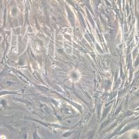
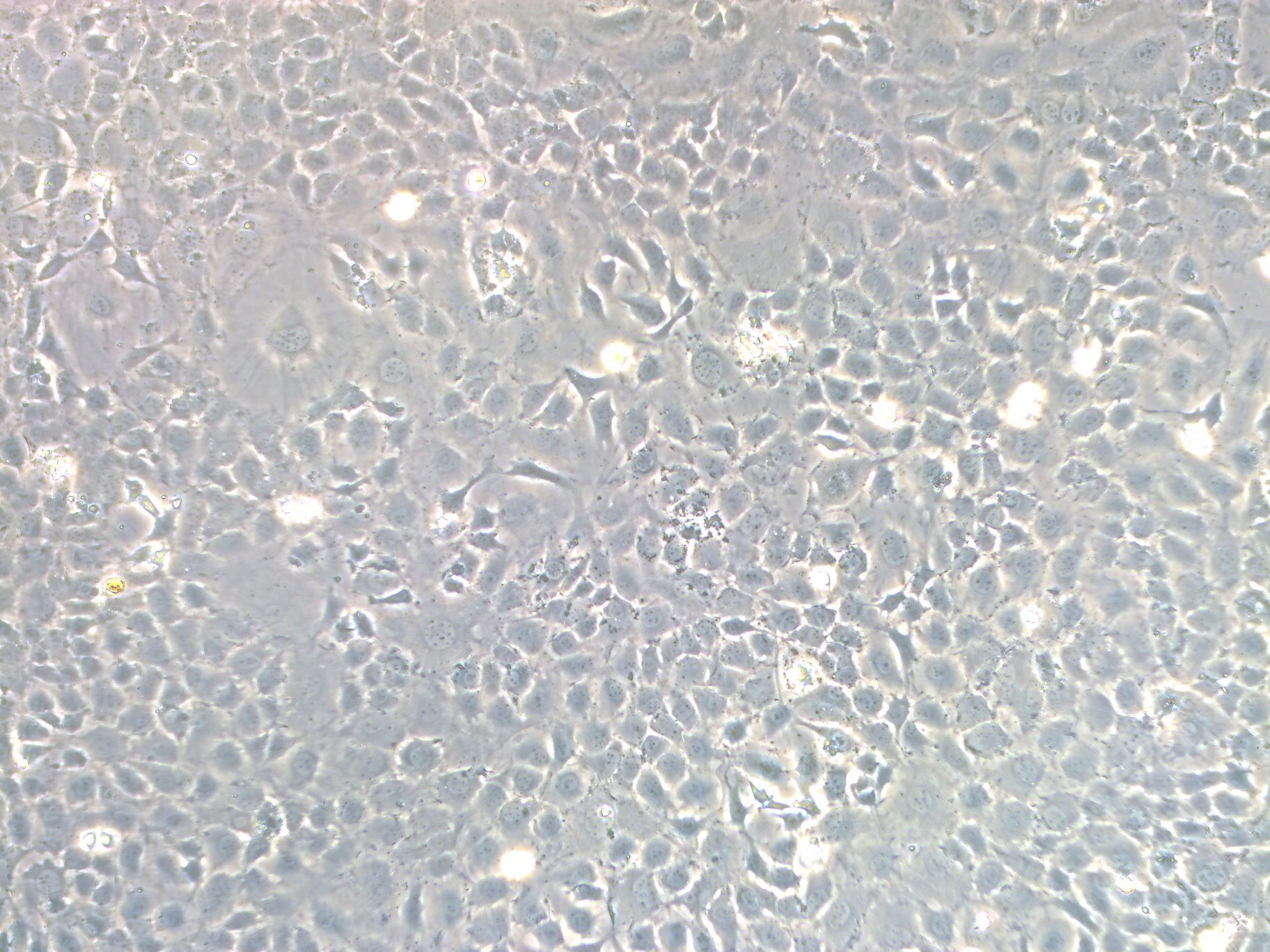
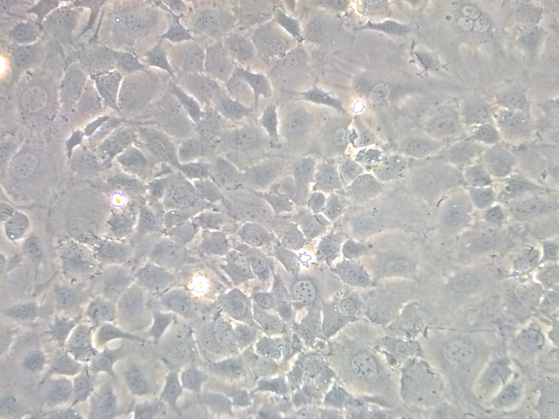
永生化小鼠软骨细胞简介:
产品描述:软骨细胞(Chondrocyte),幼稚的软骨细胞位于软骨组织的表层,单个分布,体积较小,呈椭圆形,长轴与软骨表面平行,越向深层的软骨细胞体积之间增大呈圆形,细胞核圆形或卵圆形,染色浅,细胞质弱嗜碱性,常见数量不一的脂滴。软骨细胞具有合成和分泌基质与纤维的功能。
产品货号:IM2029
产品类型: 永生化细胞
传代能力: 30代左右
产品形态:上皮型
培养基:永生化小鼠软骨细胞完全培养基
支原体:呈阴性
产品培养条件:37℃,5%CO2
发货方式:T25瓶子常温发货
货期:1周左右货期

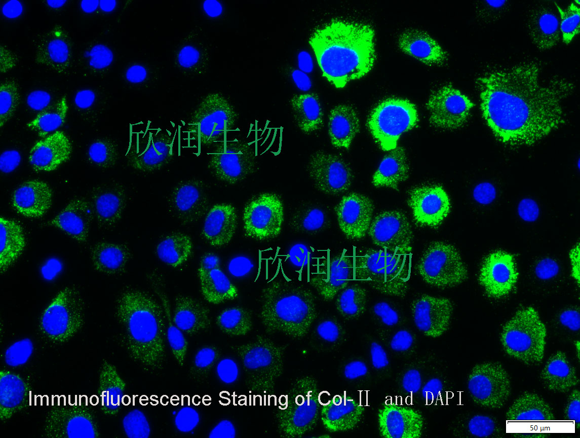
Col-II抗体免疫荧光染色鉴定


Reversibly Immortalized Mouse Articular Chondrocytes Acquire Long-Term Proliferative Capability while Retaining Chondrogenic Phenotype
Cartilage tissue engineering holds great promise for treating cartilaginous pathologies including degenerative disorders and traumatic injuries. Effective cartilage regeneration requires an optimal combination of biomaterial scaffolds, chondrogenic seed cells and biofactors. Obtaining sufficient chondrocytes remains a major challenge due to the limited proliferative capability of primary chondrocytes. Here, we investigate if reversibly immortalized mouse articular chondrocytes (iMACs) acquire long-term proliferative capability while retaining the chondrogenic phenotype. Primary mouse articular chondrocytes (MACs) can be efficiently immortalized with a retroviral vector expressing SV40 large T antigen flanked with Cre/loxP sites. iMACs exhibit long-term proliferation in culture, although the immortalization phenotype can be reversed by Cre recombinase. iMACs express the chondrocyte markers Col2a1 and aggrecan and produce chondroid matrix in micromass culture. iMACs form subcutaneous cartilaginous masses in athymic mice. Histologic analysis and chondroid matrix staining demonstrate
风险提示:丁香通仅作为第三方平台,为商家信息发布提供平台空间。用户咨询产品时请注意保护个人信息及财产安全,合理判断,谨慎选购商品,商家和用户对交易行为负责。对于医疗器械类产品,请先查证核实企业经营资质和医疗器械产品注册证情况。
 文献和实验
文献和实验Chicken intestinal epithelial cells were obtained from NEWGAINBIO company. Cells were cultured on 37℃, with 5% CO2, in the Ham’s F-12 Nutrient (DMEM/12) that contained the following supplementations: fetal bovine serum (5%), in-sulin (5 µg/mL), transferrin (5 µg/mL), selenium (5 ng/mL), epidermal growth factor (5 ng/mL) and penicillin-streptomycin (100–100 U/mL) for cell culturing (full DMEM/12). Experiments were performed with chicken intestinal epithelial cells and working solutions were prepared with plain DMEM/12 without supplementation. For the investigations, cells were seeded onto 96-well, 24-well or 6-well polystyrene cell culture plates.
Primary hVICs (passage 2) were cultured to 50–60% confluence and infected with pGMLV-SV40T-puro lentivirus (NewgainBio, Wuxi, China) at a multiplicity of infection of 80 supplemented with 5 µg/mL polybrene (Sigma-Aldrich, Buchs, Switzerland).
Tissue was cultured until cells became visible around the tissue, and when the fusion reached 90% (FIGURE 1A) §ask ¦lled with the prepared culturing medium was sent to the company for further immortalisation. Cell immortalisation was done for cell stability and longer-term use. Immortalised cells were cultured with 10% FBS and 1% PS in the DMEM medium. After the cells multiplied and merged, they were routinely passed and grown ( NEWGAINBIO Inc. Wuxi, Jiangsu, China) (FIGURE 1B-C).
Mouse primary cultured renal vascular ECs and VSMCs were obtained from Newgainbio company, which were tested by Factor VIII and α-smooth muscle actin (α-SMA), the marker of ECs and VSMCs. RNeasy Mini Kit was used for RNA extraction, and the above protocols were repeated.
Porcine primary colon epithelial cells (Newgainbio company, Wuxi,China) were cultured in Dulbecco's Modified Eagle's Medium (Solarbio, Beijing, China) containing 10 % fetal bovine serum (BioInd, Kiryat shmona, Lsrael) at 37 ◦C and 5 % CO2 humidity.
如果说动物模型告诉我们“关节发生了什么”,那么原代软骨细胞就能进一步回答:软骨细胞自己到底怎么想、怎么受伤、怎么被保护。 它不是一瓶普通细胞,而是把软骨微环境里的关键问题搬到培养皿里继续追问。 一、原代软骨细胞有什么用? 在骨关节炎、软骨损伤修复、炎症反应和基质代谢研究中,软骨细胞是绕不开的主角。 相比永生化细胞系,原代软骨细胞更接近体内细胞状态,更适合观察炎症刺激、药物干预、外泌体、材料提取液或基因调控对软骨基质的影响。 · 它能帮助判断软骨细胞是否发生炎症反应、凋亡、氧
(Finite Cell Line) : 能在体外分裂有限次数 (如人成纤维细胞系约传 50 代后停止) 后衰老的细胞。——增殖受分裂次数限制。 (2) 无限细胞系 (Immortalized Cell Lines): 指通过基因突变/转化或病毒感染,在体外培养中获得无限增殖能力的细胞群体。——通常由原代细胞转化而来,分裂次数可超过 50 代甚至无限传代。 【举例】人宫颈癌 HeLa 细胞——最著名的细胞系之一,是医学上最早经由人工培养的永生不死的细胞,被广泛用于医学研究。 图 2. HeLa
A)可诱导多能干细胞(PSC/iPSC)分化为定形内胚层(DE),随后添加 Wnt3a、成纤维细胞生长因子 4(FGF-4)和头蛋白(Noggin),可进一步引导细胞向特定谱系分化。 8.采集临床样本时如何避免污染? ①尽可能在无菌条件下进行样本采集; ②用含双抗(如青霉素 / 链霉素)的 PBS 预处理组织:对于暴露于外界环境的组织(如胃癌、肠癌、膀胱癌组织),需在含 3%-5% 双抗的 PBS 中浸泡 5-10 分钟;其他组织则使用含 1%-2% 双抗的 PBS 浸泡约 5 分钟
 技术资料
技术资料
