

- ¥4260
- 南京万木春生物
- 进口/国产
- WM-24JY599
- 2026年06月22日

企业认证
相关产品推荐更多 >
万千商家帮你免费找货
0 人在求购买到急需产品
- 详细信息
- 文献和实验
- 技术资料
- 英文名:
/
- 库存:
现货库存
- 供应商:
/
- 肿瘤类型:
/
- 细胞类型:
/
- 品系:
/
- 组织来源:
ATCC/DSMZ/ECACC
- 相关疾病:
/
- 物种来源:
人或动物
- 免疫类型:
/
- 细胞形态:
/
- 是否是肿瘤细胞:
/
- 器官来源:
/
- 运输方式:
常温或干冰
- 年限:
/
- 生长状态:
/
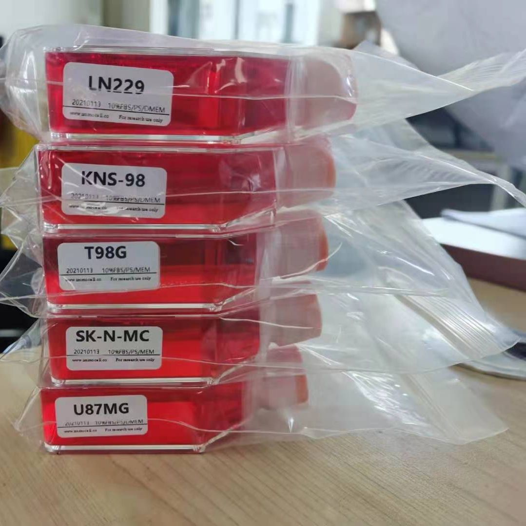
- 规格:
T25
| 种属 | 小鼠 |
| 组织来源 | 正常附睾组织 |
| 传代比例 | 1:2传代 |
| 完全培养基配置 | 基础培养基500ml ;生长添加剂5ml ;胎牛血清10ML;双抗5ml |
| 简介 | 附睾是一个多数曲折、细小的管子构成的器官 ,一面连接着输精管 ,一面连接着睾丸的曲细精管。当精子离开睾丸 时 ,就跑到附睾里 ,继续生长成熟。附睾管除贮存精子外还能分泌附睾液 ,其中含有某些激素、酶和特异的营养物 质 ,它们有助于精子的成熟。 |
with immune activities in GC. Furthermore, we demonstrated that the TP53 mutation itself could result in the depressed immune activities in GC and other cancer types. We revealed that chromosomal instability was an important mechanism for the depressed tumor immunity in employ computational methods that extract spatio-morphologic predictive features, enabling computer-aided diagnostics. We detail the benefits of computational TILs assessment, the readiness of TILs scoring for computational assessment, and outline considerations for
Objective: The objective was to assess the roles of 68Ga-PSMA PET/CT and 18F-NaF PET/CT in evaluation of skeletal metastatic lesions in prostate cancer.
Methods: Two hundred consecutive prostate cancer patients who had undergone 68Ga-PSMA PET/CT and 18F-NaF PET/CT at baseline evaluation (n = 80) and following suspected recurrence or disease progression (restaging) (n = 120) were analyzed retrospectively.
Results: PSMA and NAF scans were positive for skeletal metastatic lesions in 67% (134 patients) and negative in 33% (66 patients). The scans were concordant in 80% (160 patients: 66 negative and 94 positive) and discordant in 20% (40 patients). Among 40 discordant results, 14 were baseline and 26 were restaging studies. PSMA detected more number of lesions in 11 (nine baseline and two restaging). These were true positive marrow or lytic metastatic lesions. NaF revealed more number of lesions in 29 (5 initial and 24 restaging). These were false positive on follow-up imaging. No statistical difference (P value = 0.7 by McNemar test) between the two scans for identifying absence or presence of at least one skeletal lesion was noted at baseline staging.
风险提示:丁香通仅作为第三方平台,为商家信息发布提供平台空间。用户咨询产品时请注意保护个人信息及财产安全,合理判断,谨慎选购商品,商家和用户对交易行为负责。对于医疗器械类产品,请先查证核实企业经营资质和医疗器械产品注册证情况。
 文献和实验
文献和实验/TP53-mutated cancers. Finally, we showed that immune cell infiltration and immune activities were likely positively associated with survival prognosis in GC. Our findings suggest that p53 may play an important role in activating tumor immunity in GC and other cancer types and that the
柱状上皮主要被覆于鼻腔、鼻咽、支气管树、胃肠、子宫颈管、子宫内膜及输卵管等部位。柱状上皮脱落细胞主要包括涂片纤毛柱状细胞、粘液柱状细胞和储备细胞。 (1)纤毛柱状细胞:细胞呈锥形,顶端宽平,其表面有密集的纤毛,纤毛巴氏染色呈亮红色;胞质泡沫状,巴氏染色染蓝色,HE染淡红色;核圆形位于细胞中部,染色质细颗粒状。在涂片中的常见排列形式: 1)蜂房状排列:细胞成群或呈片医学教育|网搜集整理,排列紧密,不重叠。 2)栅栏状:细胞紧密排列,医学教育|网搜集整理可有重叠
复层鳞状上皮,一般有10多层细胞。被覆于全身皮肤、口腔、喉部、鼻咽的一部分、食道、阴道的全部以及子宫颈。鳞状上皮细胞分为基底层细胞、中层细胞和表层细胞。 (1)基底层细胞 1)内底层细胞:细胞呈圆形或卵圆形,直径12~15μm;胞质巴氏染色呈深蓝、暗绿和灰蓝色,HE染色呈暗红色;胞核圆形或卵圆形,居中,染色质细颗粒状;核与胞质比(即核的直径与细胞质幅缘之比,简称核胞质比)约1:(0.5~1)。 2)外底层细胞:细胞呈圆形或椭圆形,直径15~30μm;胞质较丰富
涂片中脱落的非上皮细胞成分又称背景成分。包括血细胞、粘液、坏死物及异特等。 1.红细胞涂片中可见到多少不等的红细胞。因红细胞大小较恒定,可作为测定其他细胞大小的标尺。红细胞量的多少与病变性质或取材时局部损伤程度有关。 2.中性粒细胞涂片中常可见多量中性粒细胞。中性粒细胞易变性,胞质溶解而成裸核。主要见于组织炎症时。此外见于癌组织坏死后继发感染时。 3.嗜酸性粒细胞其存在与炎症、变态反应或寄生虫感染有关。 4.淋巴细胞见于炎症,特别是慢性炎症时较多
 技术资料
技术资料暂无技术资料 索取技术资料

