

- ¥3500 - 4500
- 欣润生物(NEWGAINBIO)
- 江苏无锡
- IS8001
- 2026年04月26日

企业认证
相关产品推荐更多 >


万千商家帮你免费找货
0 人在求购买到急需产品
- 详细信息
- 询价记录
- 文献和实验
- 技术资料
- 英文名:
Immortalized sheep small intestinal epithelial cells
- 库存:
100万
- 供应商:
欣润生物
- 肿瘤类型:
否
- 细胞类型:
永生化
- ATCC Number:
无
- 品系:
山羊
- 组织来源:
小肠
- 相关疾病:
无
- 物种来源:
羊
- 免疫类型:
不详
- 细胞形态:
上皮型
- 是否是肿瘤细胞:
否
- 器官来源:
小肠
- 运输方式:
常温
- 年限:
/
- 生长状态:
贴壁生长
- 规格:
T25方瓶
产品介绍
产品名称:羊小肠上皮细胞系、永生化羊小肠上皮细胞
产品描述:羊小肠上皮细胞系、永生化羊小肠上皮细胞小肠是主要的消化器官,它含有肠液、胰液和胆汁,分别都含有消化蛋白质、糖、和脂肪的酶,能对蛋白质、糖、脂肪进行消化,使之变为简单的物质,如氨基酸、葡萄糖、甘油三脂等,同时胆汁能对脂肪进行分解,促进脂肪的消化。因此营养物质主要在小肠被消化,是人体的主要消化器官。而小肠上皮细胞有分泌作用,所以研究羊小肠上皮细胞系、永生化羊小肠上皮细胞对研究小肠功能有重要意义。
产品货号:IS8001
产品类型: 可传代的永生化细胞
传代能力:30代左右
产品形态: 上皮形态
培养基:羊小肠上皮细胞系、永生化羊小肠上皮细胞完全培养基
支原体检测:阴性
产品培养条件:37℃,5%CO2
发货方式:常温T25方瓶发货
货期:1周左右时间
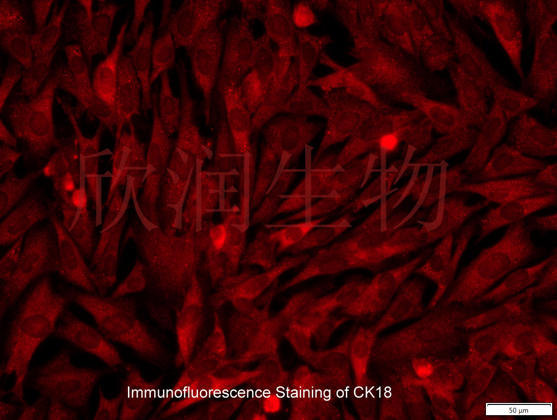
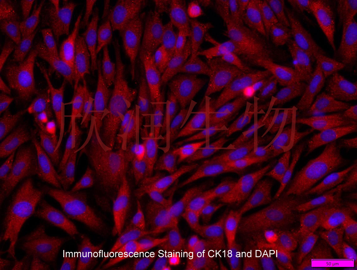
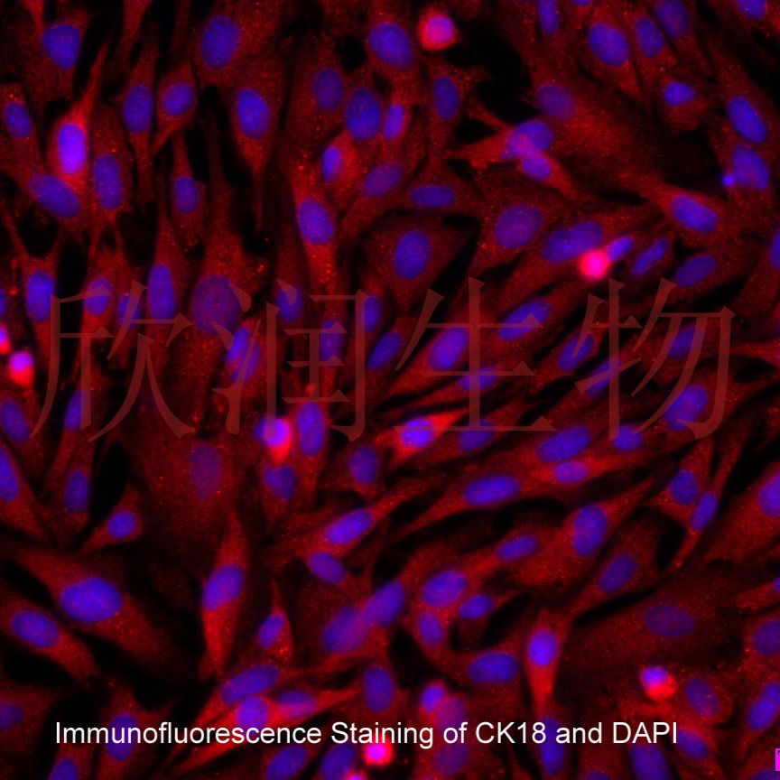
CK18免疫荧光鉴定


Generation and application of immortalized sertoli cell line from sheep testis
2023 The AuthorsPrimary sheep testicular Sertoli cells (STSCs) are ideal for investigating the molecular and pathogenic processes of capripoxvirus. However, the high cost of isolation and culture of primary STSCs, time-consuming operation, and short lifespan greatly limit their real-world application. In our study, the primary STSCs were isolated and immortalized by transfection of a lentiviral recombinant plasmid containing simian virus 40 (SV40) large T antigen. Androgen-binding protein (ABP) and vimentin (VIM) protein expression, SV40 large T antigen activity, proliferation assays, and apoptosis analysis results showed that immortalized large T antigen STSCs (TSTSCs) still had the same physiological characteristics and biological functions as primary STSCs. Moreover, immortalized TSTSCs had strong anti-apoptosis ability, extended lifespan, and enhanced proliferative activity compared to primary STSCs, which had not transformed in vitro and showed any signs of malignancy phenotype in nude mice. Besides, immortalized TSTSCs were susceptible to goatpox virus (GTPV), lumpy
风险提示:丁香通仅作为第三方平台,为商家信息发布提供平台空间。用户咨询产品时请注意保护个人信息及财产安全,合理判断,谨慎选购商品,商家和用户对交易行为负责。对于医疗器械类产品,请先查证核实企业经营资质和医疗器械产品注册证情况。
- 作者
- 内容
- 询问日期
 文献和实验
文献和实验Chicken intestinal epithelial cells were obtained from NEWGAINBIO company. Cells were cultured on 37℃, with 5% CO2, in the Ham’s F-12 Nutrient (DMEM/12) that contained the following supplementations: fetal bovine serum (5%), in-sulin (5 µg/mL), transferrin (5 µg/mL), selenium (5 ng/mL), epidermal growth factor (5 ng/mL) and penicillin-streptomycin (100–100 U/mL) for cell culturing (full DMEM/12). Experiments were performed with chicken intestinal epithelial cells and working solutions were prepared with plain DMEM/12 without supplementation. For the investigations, cells were seeded onto 96-well, 24-well or 6-well polystyrene cell culture plates.
Primary hVICs (passage 2) were cultured to 50–60% confluence and infected with pGMLV-SV40T-puro lentivirus (NewgainBio, Wuxi, China) at a multiplicity of infection of 80 supplemented with 5 µg/mL polybrene (Sigma-Aldrich, Buchs, Switzerland).
Tissue was cultured until cells became visible around the tissue, and when the fusion reached 90% (FIGURE 1A) §ask ¦lled with the prepared culturing medium was sent to the company for further immortalisation. Cell immortalisation was done for cell stability and longer-term use. Immortalised cells were cultured with 10% FBS and 1% PS in the DMEM medium. After the cells multiplied and merged, they were routinely passed and grown ( NEWGAINBIO Inc. Wuxi, Jiangsu, China) (FIGURE 1B-C).
Mouse primary cultured renal vascular ECs and VSMCs were obtained from Newgainbio company, which were tested by Factor VIII and α-smooth muscle actin (α-SMA), the marker of ECs and VSMCs. RNeasy Mini Kit was used for RNA extraction, and the above protocols were repeated.
Porcine primary colon epithelial cells (Newgainbio company, Wuxi,China) were cultured in Dulbecco's Modified Eagle's Medium (Solarbio, Beijing, China) containing 10 % fetal bovine serum (BioInd, Kiryat shmona, Lsrael) at 37 ◦C and 5 % CO2 humidity.
个雄配子,雌雄配子结合后形成合子,进入孢子生殖阶段。合子发育为卵囊,成熟的卵囊含有4个裸露的子孢子。卵囊有薄壁和厚壁两种类型。薄壁卵囊约占20%,仅有一层单位膜,其子孢子逸出后直接侵入宿主肠上皮细胞,继续无性繁殖,使宿主自身体内重复感染;厚壁卵囊约占80%,在宿主细胞或肠腔内孢子化(形成子孢子)。孢子化的卵囊随宿主粪便排出体外,即具感染性。整个生活史约需5~11天完成。用人源卵囊感染BALB/c小鼠,感染后10和11天粪检找到卵囊。 致病 隐孢子虫主要寄生于小肠上皮细胞的刷状缘
卵囊 刚从猫粪排出的卵囊为圆形或椭圆形,大小为10~12µm;具两层光滑透明的囊壁,内充满均匀小颗粒。成熟卵囊含2个孢子囊,每个分别由4个子孢子组成,相互交错在一起,呈新月形。 生活史 弓形生活史包括有性生殖和无性生殖阶段,全过程需两种宿主,在猫科动物体内完成有性世代,同时也进行无性增殖,故猫是弓形虫的终宿主兼中间宿主。在其它动物或人体内只能完成无性生殖,最为中间宿主。有性生殖只限于在猫科动物小肠上皮细胞内进行,称肠内期发育。无性生殖阶段可在肠外其它组织、细胞内进行,称肠外期
中间宿主肌肉中的肉孢子囊被终缩主吞食后,囊壁被蛋白水解酶破坏,缓殖子释出并侵入小肠固有层,无需经过裂体增殖就直接形成配子,雌雄配子交配后成为卵囊,卵囊在小肠固有层逐渐发育成熟。 致病与诊断 感染本虫后其致病作用一般不很明显,但在严重感染的急性期,即裂殖体在血管内皮细胞中增殖,释放大量裂殖子分布到全身时,可引起牛、羊的消瘦、 流产 、瘫痪和死亡。在肌肉中的肉孢子囊可破坏所侵犯的肌细胞,当长大时可造成邻近细胞的压迫性萎缩,如囊壁破裂
 技术资料
技术资料
