大家都在搜

手机验证

询价列表

暂时没有已询价产品

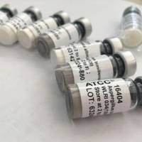
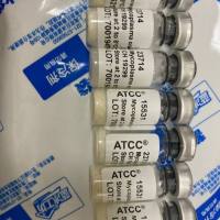
ATCC vEPT细胞

企业认证
vEPT
大量
北京百奥创新科技有限公司
上皮成纤维细胞
肾;近端小管
正常
Oryctolagus cuniculus;兔子
上皮细胞样
是
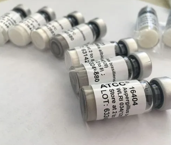
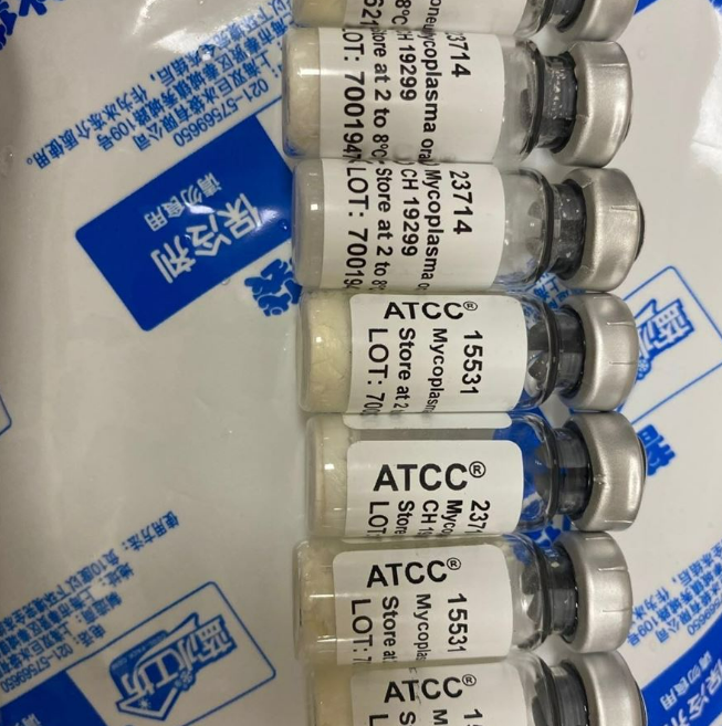
产品信息:
| 货号 | 产品名称 | 规格 |
| CRL-2087 | vEPT | 支 |
风险提示:丁香通仅作为第三方平台,为商家信息发布提供平台空间。用户咨询产品时请注意保护个人信息及财产安全,合理判断,谨慎选购商品,商家和用户对交易行为负责。对于医疗器械类产品,请先查证核实企业经营资质和医疗器械产品注册证情况。
 技术资料
技术资料