


明星产品※美天旎Miltenyi货号130-051-301现
货human CD138 MicroBeads上海睿安生物19121878899美天旎Miltenyi官方正式授权代理商- ¥9102.15
- 美天旎Miltenyi
- 130-051-301
- Made in Germany
- 2026年04月26日

企业认证
相关产品推荐更多 >
![Miltenyi货号130-096-533现货CD4⁺ T Cell Isolation Kit(human)[CD4⁺ T细胞分离试剂盒]13611631389上海睿安生物](https://img1.dxycdn.com/p/s14/2025/0529/525/0694628836252697291.jpg!wh200)
Miltenyi货号130-096-533现货CD4⁺ T Cell Isolation Kit(human)[CD4⁺ T细胞分离试剂盒]13611631389上海睿安生物
¥11826.58![美天旎Miltenyi货号130-117-043小鼠CD4(L3T4)磁珠[mouse CD4(L3T4) MicroBeads]上海睿安生物13611631389](https://img1.dxycdn.com/p/s14/2025/0621/473/4492255803412446391.jpg!wh200)
美天旎Miltenyi货号130-117-043小鼠CD4(L3T4)磁珠[mouse CD4(L3T4) MicroBeads]上海睿安生物13611631389
¥9149.61
美天旎Miltenyi货号130-097-052人CD14磁珠CD14 MicroBeads(human-lyophilized)上海睿安生物13611631389
¥8030.91
美天旎Miltenyi货号130-090-101死细胞去除试剂盒Dead Cell Removal Kit上海睿安生物13611631389
¥4992.34
Miltenyi货号130-045-201人CD8磁珠(human CD8 MicroBeads)上海睿安生物13611631389
¥8030.91
万千商家帮你免费找货
0 人在求购买到急需产品
- 详细信息
- 文献和实验
- 技术资料
- 英文名:
human CD138 MicroBeads
- 保质期:
1年
- 保存条件:
2-8°C
- 库存:
10⁺盒
- 供应商:
上海睿安生物19121878899
- 规格:
2✕10⁹ total cells/盒
明星产品※美天旎Miltenyi货号130-051-301现货human CD138 MicroBeads上海睿安生物19121878899美天旎Miltenyi官方正式授权代理商
CD138 MicroBeads were developed for the positive selection or depletion of plasma cells from PBMCs, bone marrow, leukapheresis harvests, and single-cell suspensions from lymphoid tissues.
Overview
CD138 MicroBeads were developed for the positive selection or depletion of plasma cells from PBMCs, bone marrow, leukapheresis harvests, and single-cell suspensions from lymphoid tissues.
Detailed product information
The CD138 antigen, also known as syndecan-1, is primarily expressed on normal and malignant plasma cells in the bone marrow. It is further found on a subset of plasma cells in peripheral blood and certain lymphoid tissues. The CD138 antigen is neither expressed on naive B cells, germinal center B cells, memory B cells, nor on T cells or monocytes.
Since the direct study of plasma cells is hampered by their low frequency, prior enrichment of plasma cells using CD138 MicroBeads allows more accurate cell analysis, e.g. by flow cytometry2. To ease the analysis of enriched plasma cells, CD138 MicroBeads are also available in combination with CD138-PE. Plasma cells purified with CD138 MicroBeads are applied for molecular biological studies, such as PCR1,4,7,8, microarrays2,4,5,6, or protein analysis7.
For positive selection: MS, LS, XS, or autoMACS® Columns. For depletion: LD, D, or autoMACS Columns.
Figure 1
CD138+ plasma cells were isolated from normal PBMCs using CD138 MicroBeads, an MS Column, and a MiniMACS™ Separator.
-
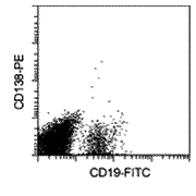 PBMCs before separation
 -
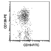 Enriched CD138+ cells
Citations for CD138 MicroBeads, human
Publications
Horst, A. et al. (2002) Detection and characterization of plasma cells in peripheral blood: correlation of IgE+ plasma cell frequency with IgE serum titre. Clin. Exp. Immunol. 130: 370-378
Draube, A. et al. (2001) Immunomagnetic enrichment of CD138 positive cells from weakly infiltrated myeloma patients samples enables the determination of the tumor clone specific IgH rearrangement. Ann. Hematol. 80: 83-89
Lee, J. W. et al. (2004) IL-3 expression by myeloma cells increases both osteoclast formation and growth of myeloma cells. Blood 103: 2308-2315
Kumar, S. et al. (2004) Bone marrow angiogenic ability and expression of angiogenic cytokines in myeloma: evidence favoring loss of marrow angiogenesis inhibitory activity with disease progression. Blood 104: 1159-1165
Davies, F. E. et al. (2003) Insights into the multistep transformation of MGUS to myeloma using microarray expression analysis. Blood 102: 4504-4511
Zhan, F. et al. (2003) Gene expression profiling of human plasma cell differentiation and classification of multiple myeloma based on similarities to distinct stages of late-stage B-cell development. Blood 101: 1128-1140
Tarte, K. et al. (2003) Gene expression profiling of plasma cells and plasmablasts: toward a better understanding of the late stages of B-cell differentiation. Blood 102: 592-600
Shaughnessy, J. Jr. et al. (2001) Cyclin D3 at 6p21 is dysregulated by recurrent chromosomal translocations to immunoglobulin loci in multiple myeloma. Blood 98: 217-223

风险提示:丁香通仅作为第三方平台,为商家信息发布提供平台空间。用户咨询产品时请注意保护个人信息及财产安全,合理判断,谨慎选购商品,商家和用户对交易行为负责。对于医疗器械类产品,请先查证核实企业经营资质和医疗器械产品注册证情况。
 文献和实验
文献和实验明星产品※美天旎Miltenyi货号130-051-301现货human CD138 MicroBeads上海睿安生物19121878899美天旎Miltenyi官方正式授权代理商
CD138 MicroBeads were developed for the positive selection or depletion of plasma cells from PBMCs, bone marrow, leukapheresis harvests, and single-cell suspensions from lymphoid tissues.
Overview
CD138 MicroBeads were developed for the positive selection or depletion of plasma cells from PBMCs, bone marrow, leukapheresis harvests, and single-cell suspensions from lymphoid tissues.
Detailed product information
The CD138 antigen, also known as syndecan-1, is primarily expressed on normal and malignant plasma cells in the bone marrow. It is further found on a subset of plasma cells in peripheral blood and certain lymphoid tissues. The CD138 antigen is neither expressed on naive B cells, germinal center B cells, memory B cells, nor on T cells or monocytes.
Since the direct study of plasma cells is hampered by their low frequency, prior enrichment of plasma cells using CD138 MicroBeads allows more accurate cell analysis, e.g. by flow cytometry2. To ease the analysis of enriched plasma cells, CD138 MicroBeads are also available in combination with CD138-PE. Plasma cells purified with CD138 MicroBeads are applied for molecular biological studies, such as PCR1,4,7,8, microarrays2,4,5,6, or protein analysis7.
For positive selection: MS, LS, XS, or autoMACS® Columns. For depletion: LD, D, or autoMACS Columns.
Figure 1
CD138+ plasma cells were isolated from normal PBMCs using CD138 MicroBeads, an MS Column, and a MiniMACS™ Separator.
-
PBMCs before separation
-
Enriched CD138+ cells
Citations for CD138 MicroBeads, human
Publications
Horst, A. et al. (2002) Detection and characterization of plasma cells in peripheral blood: correlation of IgE+ plasma cell frequency with IgE serum titre. Clin. Exp. Immunol. 130: 370-378
Draube, A. et al. (2001) Immunomagnetic enrichment of CD138 positive cells from weakly infiltrated myeloma patients samples enables the determination of the tumor clone specific IgH rearrangement. Ann. Hematol. 80: 83-89
Lee, J. W. et al. (2004) IL-3 expression by myeloma cells increases both osteoclast formation and growth of myeloma cells. Blood 103: 2308-2315
Kumar, S. et al. (2004) Bone marrow angiogenic ability and expression of angiogenic cytokines in myeloma: evidence favoring loss of marrow angiogenesis inhibitory activity with disease progression. Blood 104: 1159-1165
Davies, F. E. et al. (2003) Insights into the multistep transformation of MGUS to myeloma using microarray expression analysis. Blood 102: 4504-4511
Zhan, F. et al. (2003) Gene expression profiling of human plasma cell differentiation and classification of multiple myeloma based on similarities to distinct stages of late-stage B-cell development. Blood 101: 1128-1140
Tarte, K. et al. (2003) Gene expression profiling of plasma cells and plasmablasts: toward a better understanding of the late stages of B-cell differentiation. Blood 102: 592-600
Shaughnessy, J. Jr. et al. (2001) Cyclin D3 at 6p21 is dysregulated by recurrent chromosomal translocations to immunoglobulin loci in multiple myeloma. Blood 98: 217-223


