

- 询价
- TheWell

- VHM01
- 2025年11月24日

企业认证
相关产品推荐更多 >

Dulbecco's Modified Eagle Medium (DMEM), w/o D-Glucose, w/o Sodium Pyruvate, w/o L-Glutamine DMEM
询价Schneider's Drosophila Medium, with L-Glutamine 施耐德的果蝇培养基(含谷氨酰胺)
询价VC-ECL Kit HRP化学发光增强试剂盒
询价
瑞典BioLamina人类重组层粘连蛋白Laminin-211,LN211
询价Minimum Essential Medium (MEM), Earle's Salts Base, with Non-Essential Amino Acids, w/o L-Glutamine MEM(含非必需氨基酸NEAA,不含谷氨酰胺)
询价
万千商家帮你免费找货
0 人在求购买到急需产品
- 详细信息
- 询价记录
- 技术资料
Overview
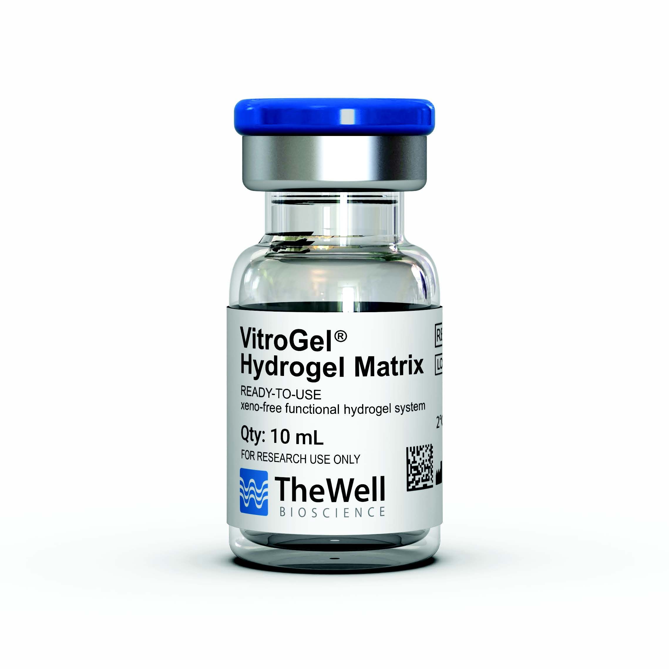
VitroGel® Hydrogel Matrix is a ready-to-use, xeno-free functional hydrogel for 3D cell culture research. VitroGel Hydrogel Matrix is an optimized formulation of multi-functional ligands and concentration to support a wide range of cell types for different applications.
“Just Add Cells” – The hydrogel matrix is ready to mix with cell suspension directly. There is no additional adjustment needed.
VitroGel Hydrogel Matrix closely mimics the natural extracellular matrix (ECM) environment to make cells feel more like at home. The hydrogel is room temperature stable, has a neutral pH, transparent, permeable and compatible with different imaging systems. The solution transforms into a hydrogel matrix by simply mixing with the cell culture medium. Cells cultured in this system can be easily harvested out with our VitroGel Cell Recovery Solution.
This user-friendly functional hydrogel creates an excellent balance of simplicity and versatility.
Specifications
| Formulation | Xeno-free, functional hydrogel |
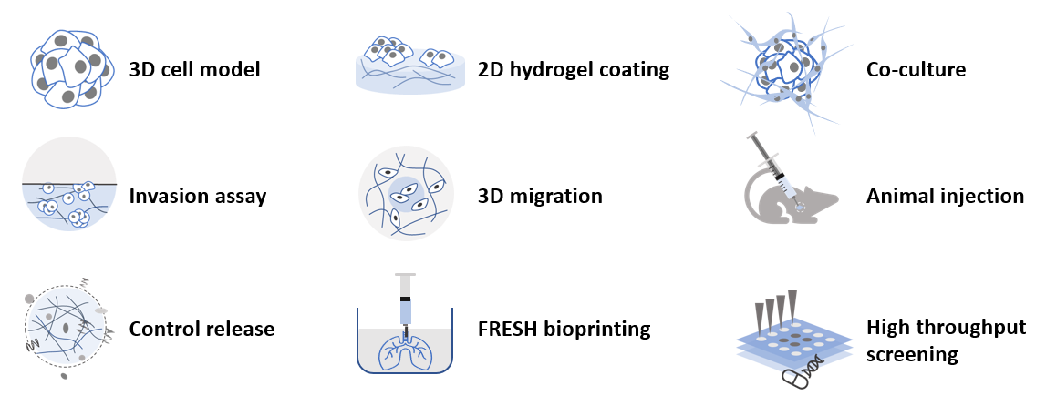
| Use | 3D and 2D cell culture |
| Operation | Ready-to-use at room temperature |
| Biocompatibility | Biocompatible, safe for animal studies |
| Injection | Injectable hydrogel for in vivostudies and lab automation |
| Cell Harvesting | 20 min cell recovery using VitroGel Cell Recovery Solution |
| pH | Neutral |
| Storage | Store at 2-8°C. Ships at ambient temperature |
| Sizes | 10 mL and 2 mL |
| Usage | 300 uses (10 mL), 60 uses (2mL) |
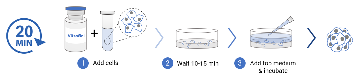
3D cell culture process in 20 min – “Just add cells”
VitroGel Hydrogel Matrix is ready-to-use. Just mix with your cells. There is no cross-linking agent or the need to adjust the hydrogel concentration.
Handbooks and Resources
Product Documentation
Data and References
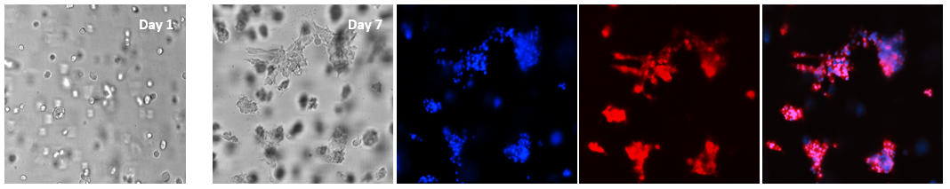
Figure 1. 3D culture of glioblastoma cells (U-87 MG) in VitroGel Hydrogel Matrix
The single-cell suspension was prepared with 50% FBS and mixed with VitroGel Hydrogel Matrix at 4:1 v/v ratio (400 µL VitroGel + 400 µL cell suspension) for the final 10% FBS in the mixture. The cells were cultured in the hydrogel matrix for 7 days and stained with live-cell imaging dyes. The nucleic acid dye (Hoechst 33342, Blue) and cell membrane dye (Red) were added directly in the cover medium and incubated for 30 minutes at 37°C before imaging. Z-stack imaging system with 2D image projection was used for both transmitted light and fluorescent images. The glioblastoma cells grew in the hydrogel matrix and formed 3D colonies. The cell-cell interactions were showed in the extended structure that connected different colonies. The VitroGel Hydrogel Matrix shows great support for 3D glioblastoma cell models with strong cell-cell communications. The hydrogel is extremely easy for image analysis, e.g, by adding the molecular probes directly on the top of the hydrogel.
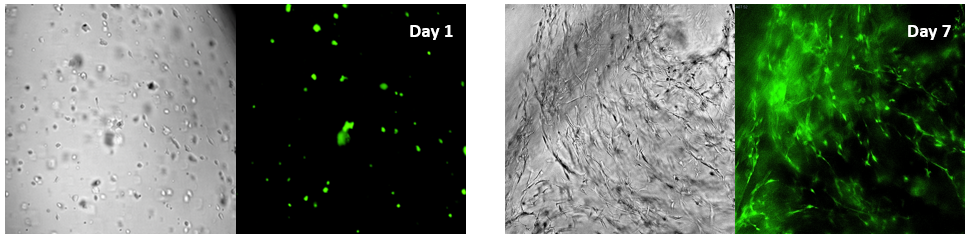 Figure 2. Bone marrow cells 3D cultured in VitroGel Hydrogel Matrix
The fibroblast-like mouse bone marrow stromal cells (OP9-GFP) were 3D cultured in VitroGel Hydrogel Matrix. The single cells were homogenously suspended within the hydrogel matrix and started to form the stretched fibroblast-like structure on day 1. The multiple integrin-binding ligands and cellular functional ligands on VitroGel Hydrogel Matrix can promote a strong cell-matrix interaction, which accelerated the cell proliferation and cell-cell communications during the 3D cell culture. On day 7, a clear 3D cellular networking structure formed within the hydrogel matrix.Z-stack imaging system with 2D image projection were used for both transmitted light and GFP fluorescent images.
Figure 3. Cancer cells grown in ViroGel Hydrogel Matrix
Various cancer cells 3D cultured in VitroGel Hydrogel Matrix can grow rapidly and form tumor-like structures. The images above show human mammary breast cancer cells (MCF-7), human prostatic cancer cells (PC3) and human pancreatic cancer cells (PANC-1) 3D cultured in VitroGel Hydrogel Matrix. The cells were prepared as single cells suspension and encapsulated within the hydrogel matrix. The grape-like shaped cell colonies in MCF-7 cells starts to form (day 1). The cell colonies continue to grow and formed the tumor-like structure (day 7). A similar phenomenon was also presented in PC3 and PANC-1 cells where the cells grew rapidly into a 3D mini-tumor within the hydrogel matrix.
风险提示:丁香通仅作为第三方平台,为商家信息发布提供平台空间。用户咨询产品时请注意保护个人信息及财产安全,合理判断,谨慎选购商品,商家和用户对交易行为负责。对于医疗器械类产品,请先查证核实企业经营资质和医疗器械产品注册证情况。
- 作者
- 内容
- 询问日期
 技术资料
技术资料暂无技术资料 索取技术资料
