Imaging the Spatial Orientation of Subunits Within Membrane Receptors by Atomic Force Microscopy
互联网
互联网
相关产品推荐

大鼠 PPAR-α (Peroxisome Prolife大鼠ors Activator Receptors alpha) ELISAKit
¥960

TAF12/TAF12蛋白Recombinant Human Transcription initiation factor TFIID subunit 12 (TAF12)重组蛋白Transcription initiation factor TFIID 20/15KDA subunits ;TAFII-20/TAFII-15 ;TAFII20/TAFII15蛋白
¥1344

IL-5R alpha重组蛋白|Recombinant Human IL-5R alpha Protein (Membrane-bound, His Tag)
¥3480

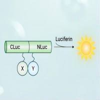
荧火素酶互补实验(Luciferase Complementation Assay, LCA)| 荧光素酶互补成像技术(Luciferase Complementation Imaging, LCI)
¥5999

Rat PPAR-α (Peroxisome ProlifeRators Activator Receptors alpha) ELISA试剂盒
¥960

