大家都在搜

手机验证

询价列表

暂时没有已询价产品
HSP70 High Sens
itivity ELISA kit

企业认证
3-4周
博士德生物
酶联免疫双抗夹心法
cell lysates,Plasma,serum,Tissue
Mouse
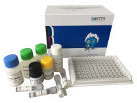
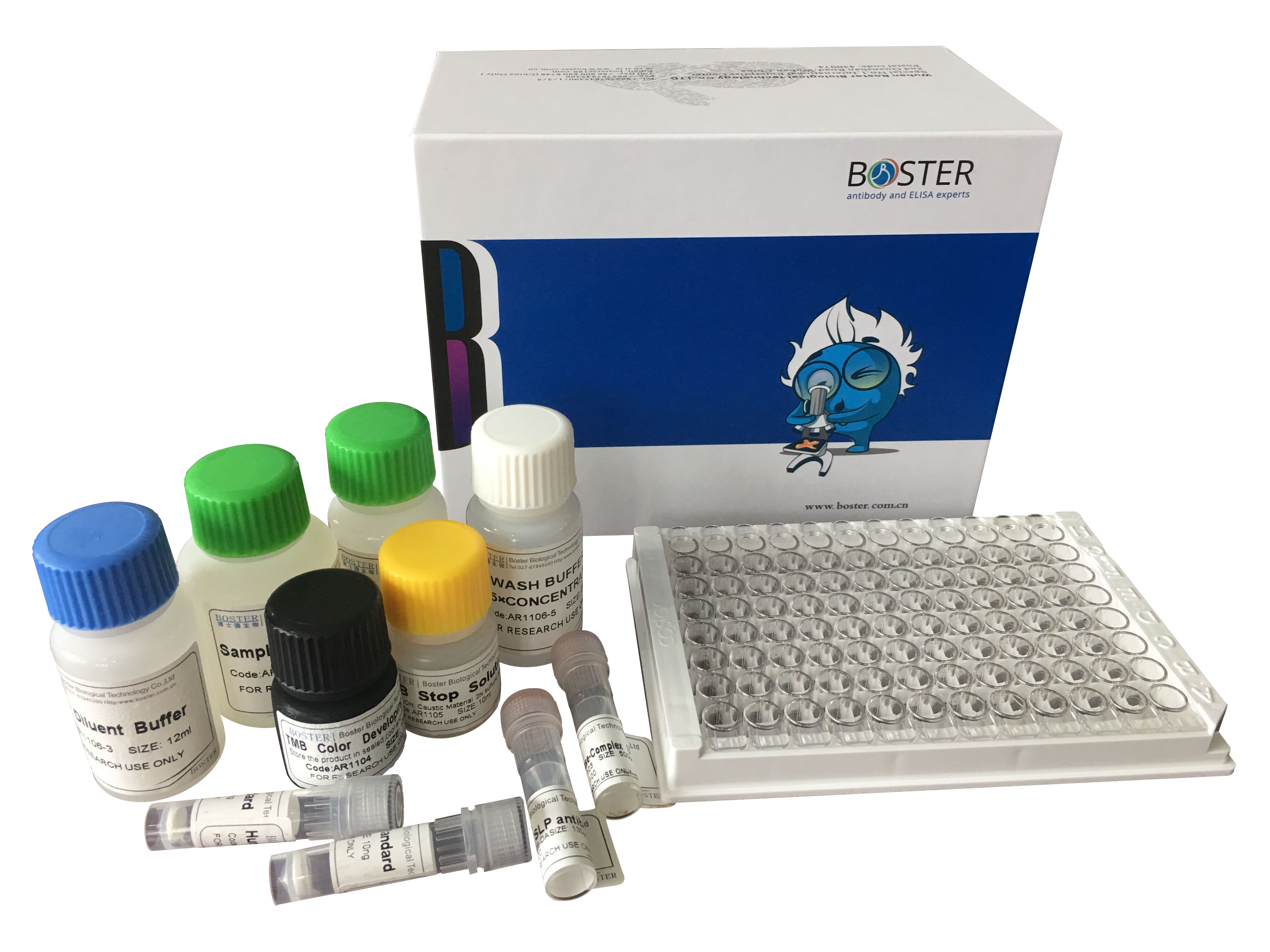
96T
This ELISA kit is of sandwich format. A sandwich ELISA measures antigen between two layers of antibodies (capture and detection antibody). After an incubation period, any unbound antibody is washed off. Enzyme substrate (for example, TMB for HRP) is added to each well and will be transformed into a blue precipitate, the amount of which is linearly proportional to the amount of enzyme in the well. The precipitate is then turned into yellow by adding the acid stop solution and the concentration of yellow precipitate is read at 450nm for light absorbance (O.D. value). The O.D. is then used to calculate the amount of molecule of interest in each well, by comparing each sample well against the standard curve. The standard curve is generated using the same principle but instead of adding samples, a series of recombinant molecules with known concentrations are added to 6-8 wells.
| Product Name | HSP70 High Sensitivity ELISA kit |
|---|---|
| SKU/Catalog Number | EK7009 |
| Description | Sandwich High Sensitivity ELISA kit for Quantitative Detection of HSP70. 96wells/kit |
| Cite This Product | HSP70 High Sensitivity ELISA kit (Boster Biological Technology, Pleasanton CA, USA, Catalog # EK7009) |
| Validated Species | Mouse |
| Application | ELISA *Our Boster Guarantee covers the use of this product in the above tested applications. |
| Cross Reactivity | There is no detectable cross-reactivity. |
| Pack Size | 96wells/kit, with removable strips. |
| Sensitivity | 0.02 ng/ml *Sensitivity, or Lower Limit of Detection (LLD), is the minimum level of target protein the ELISA assay can detect. We measure 20 blank wells and if the O.D. value is 2 standard deviations higher than the blanks' average O.D. the sample can be deemed positive. |
|---|---|
| Assay Range | 0.55 - 35 ng/ml *This assay range is determined using common samples. For samples with low target protein concentrations, users can adjust the standard curve to extend the lower limit of assay range. |
| Sample Type | Cell lysates, Plasma, Serum, Tissue *The above listed samples are the ones valided with the assay. If you do not see your sample of interest listed, as long as there is enough level of target protein present in the sample, this Picokine? ELISA kit should detect it. **For protocol and tips regarding preparing your sample of interest, please check our ELISA sample preparation guide. |
| Storage | Store the kit at 4°C. The reagents are stable until expiration of the kit. Do not expose reagent to heat, sun, or strong light. Avoid multiple freeze-thaw cycles(Shipped with wet ice.) |
| Description | Quantity |
| Anti-Hsp70 Immunoassay Plate | 12x8x1 Microwells |
| Recombinant Hsp70 Standard | 2 vials |
| Standard and Sample Diluent | 1 vial/ 50 ml |
| 10X Wash Buffer Concentrate | 1 vial/100 ml |
| Anti-Hsp70 Biotinylated Antibody Concentrate | 1 vial/150 ?l |
| Anti-Hsp70 Biotinylated Antibody Diluent | 1 vial/ 13 ml |
| Streptavidin: HRP Concentrate | 1 vial/50 ?l |
| Streptavidin: HRP Diluent | 1 vial/ 13 ml |
| TMB Substrate | 1 vial/ 13 ml |
| Stop Solution | 1 vial/ 13 ml |
1. Distilled or deionized water
2. Precision pipettes
3. Disposable pipette tips. Multichannel pipettes are recommended in the condition of large amount of samples in the detection.
4. ELISA plate reader capable of measuring at 450nm
HSP70 genes encode abundant heat-inducible 70-kDa HSPs (HSP70s). In most eukaryotes HSP70 genes exist as part of a multigene family. They are found in most cellular compartments of eukaryotes including nuclei, mitochondria, chloroplasts, the endoplasmic reticulum and the cytosol, as well as in bacteria. The genes show a high degree of conservation, having at least 5O% identity. The N-terminal two thirds of HSP70s are more conserved than the C-terminal third. HSP70 binds ATP with high affinity and possesses a weak ATPase activity which can be stimulated by binding to unfolded proteins and synthetic peptides. When HSC70 (constitutively expressed) present in mammalian cells was truncated, ATP binding activity was found to reside in an N-terminal fragment of 44kDa which lacked peptide binding capacity. Polypeptide binding ability therefore resided within the C-terminal half. The structure of this ATP binding domain displays multiple features of nucleotide binding proteins. All HSP70s, regardless of location, bind proteins, particularly unfolded ones. The molecular chaperones of the HSP70 family recognize and bind to nascent polypeptide chains as well as partially folded intermediates of proteins preventing their aggregation and misfolding. The binding of ATP triggers a critical conformational change leading to the release of the bound substrate protein. The universal ability of HSP70s to undergo cycles of binding to and release from hydrophobic stretches of partially unfolded proteins determines their role in a great variety of vital intracellular functions such as protein synthesis, protein folding and oligomerization and protein transport.
Click the images to enlarge.
[list_product_images] | [/list_product_images]
风险提示:丁香通仅作为第三方平台,为商家信息发布提供平台空间。用户咨询产品时请注意保护个人信息及财产安全,合理判断,谨慎选购商品,商家和用户对交易行为负责。对于医疗器械类产品,请先查证核实企业经营资质和医疗器械产品注册证情况。