

- 询价
- Nikon
- C2
- 日本
- 2025年07月14日


万千商家帮你免费找货
0 人在求购买到急需产品
- 详细信息
- 询价记录
- 文献和实验
- 技术资料
- 库存:
大量
- 规格:
套
An essential microscopy laboratory instrument
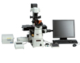
Configured with Ni-E
The C2+ confocal microscope system is part of a new generation of Nikon confocal instruments designed to be essential laboratory microscopy tools. Built on a reputation of incredible stability and operational simplicity coupled with superior optical technologies and high-speed image acquisition of up to 100 fps*, the C2+ is the perfect tool for a new microscope, or as a new accessory to an existing Nikon imaging system.
- *With 8x zoom or larger
Large field-of-view imaging and three-dimensional reconstruction
Confocal image acquisition, using high-numerical aperture and high-magnification objectives, together with XY stage control and advanced image stitching with Nikon NIS-Elements software, enables high-resolution images of large areas of a specimen to be produced. In addition, the microscope's high-precision Z-axis control allows assemblage of Z stack images for three-dimensional image reconstruction.
Image Stitching (Large Image)
Specimen: Genital tract of Drosophila melanogaster
Photo courtesy of: Director and Professor Masatoshi Yamamoto, Drosophila Genetic Resource Center, Kyoto Institute of Technology
Image Quality
Nikon's unprecedented optics and a time-proven, highly efficient optical design provide the brightest and sharpest images, at the longest working distances.
High-efficiency scanning heads and detectors
With the convenient, small scan head size, the C2+ can be used with various types of Nikon microscope. The C2+ employs high precision mirrors and optically superior circular pinholes, and separates the detectors to isolate sources of heat and noise, enabling low-noise, high-contrast and high-quality confocal imaging. The newly developed scanner driving system and Nikon's unique image correction technique allow 8 fps (512 x 512 pixels) and 100 fps (512 x 32 pixels) high-speed imaging.
High-performance optics
CFI Apochromat 40xWI λS, NA1.25 (left)
CFI Apochromat LWD 40xWI λS, NA1.15 (middle)
CFI Apochromat 60x Oil λS, NA1.4 (right)
CFI Apochromat λS Series
These high-numerical aperture (NA) objectives are ideal for confocal imaging with correction of chromatic aberrations over a wide wavelength range from ultraviolet. In particular, the LWD 40xWI lens corrects up to infrared. Transmission is increased through the use of Nikon's exclusive Nano Crystal Coat technology.
CFI Apochromat TIRF Series
CFI Apochromat TIRF 60x oil, NA1.49 (left)
CFI Apochromat TIRF 100x oil, NA1.49 (right)
These objectives boast an unprecedented NA of 1.49 (using a standard coverslip and immersion oil), the highest resolution among Nikon objectives. The temperature correction ring corrects image quality affected by temperature change in the range of 23°C to 37°C.
High-definition diascopic DIC images
The C2+ can acquire simultaneous three-channel fluorescence or simultaneous three-channel and diascopic DIC observation. High-quality DIC images and fluorescence images can be superimposed to aid in morphological analysis.
DIC image
Overlay of DIC and fluorescence images
High functionality
High-performance imaging software NIS-Elements offers a variety of image processing and analysis functions. It also enables data extraction from acquired images. In addition, NIS-Elements allows for intuitive operation of Nikon microscopes and other third-party peripheral devices, such as EMCCD cameras and filter wheels, to broaden the range of experiments possible.
Multimode capability
Various imaging methods, such as confocal, widefield, TIRF, photoactivation, as well as processing, analysis and presentation of acquired images, are available in one software package. Users can easily learn how to control different imaging systems with a common interface and workflow.
Easy-to-recognize display for setting lasers, detectors, etc.
Scanning parameter settings
Unmixing
Spectral analysis GUI
Numerous functions for analysis and unmixing of acquired spectrums are provided, while spectral profiles of general dyes and fluorescent proteins are preprogrammed.
Flexibility
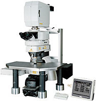
The C2+ can be coupled with upright, inverted, physiological, and macro imaging microscopes and has options for combinations with various high-quality research experiment systems. All can be controlled with NIS-Elements software.
TIRF/Photoactivation-C2+ Multimode imaging system
Optional TIRF laser illumination module and a photoactivation module can be integrated to enable both imaging of single molecules with an extremely high S/N ratio, and imaging of the fluorescence characteristic changes of photoactivated and photo-convertible fluorescent protein.
AZ-C2+ Macro confocal microscope system
With a high-definition large field of view, specimens larger than 1cm can be acquired with an unprecedentedly high S/N ratio. The AZ-C2+ allows for imaging of whole-mount specimens, such as embryos, in a single acquisition, up to 4000x4000 pixel resolution, and it can also acquire 32-channel spectral data with the C2si+. It offers a combination of low and high magnification objective lenses, optical zoom and a confocal scanning zoom function, enabling continuous imaging from macro to micro.
TT2 ES cells
Anti-Nanog antibody (Cy3), anti-Oct3/4 antibody (Alexa488) and DAPI localized in cell nuclei
Photographed with the cooperation of: Hiroshi Kiyonari, Laboratory for Animal Resources and Genetic Engineering, RIKEN Center for Developmental Biology
Photo courtesy of: Director and Professor Masatoshi Yamamoto, Drosophila Genetic Resource Center, Kyoto Institute of Technology
Some sample images in this brochure were captured using the C1 confocal microscope system.
风险提示:丁香通仅作为第三方平台,为商家信息发布提供平台空间。用户咨询产品时请注意保护个人信息及财产安全,合理判断,谨慎选购商品,商家和用户对交易行为负责。对于医疗器械类产品,请先查证核实企业经营资质和医疗器械产品注册证情况。
- 作者
- 内容
- 询问日期
 文献和实验
文献和实验相关专题 科研推动力:荧光显微镜 尼康推出的新型TE2000系列倒置显微镜 超越时代步伐,适用于所有活细胞的先进应用,为的孕育诞生为活细胞研究提供了顶尖水平的光学成像,为活细胞多维成像提供了理想平台。CFI60光学系统确保了镜头的长工作距离和高数值孔径;新型DIC系统清晰再现了标本的微小结构,包括三种DIC棱镜可供选择:标准型、高衬度型、高分辨率型;所有机型都可以选配电动功能。 多端口、分层结构 设计
用其他更高级的荧光显微镜呢?armstrong:当然可以观察的到,不过你没事观察细胞自发荧光干什么?应该尽量减少自发光。只要你的荧光显微镜配置的滤光片的波长范围把488nm和507nm(EX and Em,respectively)罩住即可了。yjr923:请教奥林帕斯与尼康哪个品牌的显微镜更好一些?llhawk:推荐使用NIKON或者LEICA的显微镜。特别是LEICA的显微镜,小巧,方便,性能好。goldenearth:我欲做卵母细胞培养急需体视显微镜,不知国产的那个牌子比较好?20,40倍,不需
的FLUAR 20×物镜。 当然还有其他更特殊的物镜,比如金相显微镜用的无盖玻片物镜和反射暗视野物镜、超低倍物镜等。 5、物镜上有很多的参数标记,来帮助大家识别物镜的等级和功能。 下面用NIKON的一款物镜做例子以解释物镜的重要参数: (1) NIKON ——制造商名称:尼康公司 (2) PLAN APO——物镜的名称:平场复消色差物镜 (3) 60× 放大倍数:60倍 (4) 0.95 数值孔径:0.95 (6) DIC M 功能
 技术资料
技术资料暂无技术资料 索取技术资料

