

- 询价
- YSKD
- 大鼠雾化仪
- 北京
- 2026年05月02日
企业认证

万千商家帮你免费找货
0 人在求购买到急需产品
- 详细信息
- 文献和实验
- 技术资料
- 保修期:
12个月
- 现货状态:
现货
药效学评价:在疾病模型中验证候选药物的治疗效果。
药代动力学研究:精准评估药物经肺部的吸收、分布和代谢特性。
毒理学研究:考察新药对呼吸系统的潜在刺激性或毒性,确保安全性。
4. 提升数据质量与可重复性
相比侵入性操作(如气管滴注),雾化给药能大幅减少动物的应激反应,避免应激对免疫和内分泌系统的干扰。确保了实验的高度可重复性和数据的准确性,符合动物伦理和科学规范。
5. 支持前沿领域探索
该仪器也是拓展肺部基因治疗、核酸药物递送和纳米靶向制剂等前沿研究的关键平台,用于评估新型治疗方式在肺部的沉积、渗透与效果。
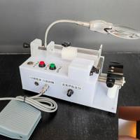
(二)北京元森凯德研制生产,药学专用小动物雾化给药仪性能优势:
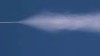
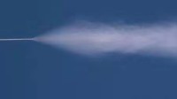
①精准可控的气溶胶发生:
采用先进的双流体雾化技术,可生成高度均一、稳定的气溶胶颗粒。MMAD 通常在 1-5 μm 范围内,确保药物能高效沉积于实验动物的特定呼吸道区域(如肺泡或支气管),完美模拟人体吸入给药过程。
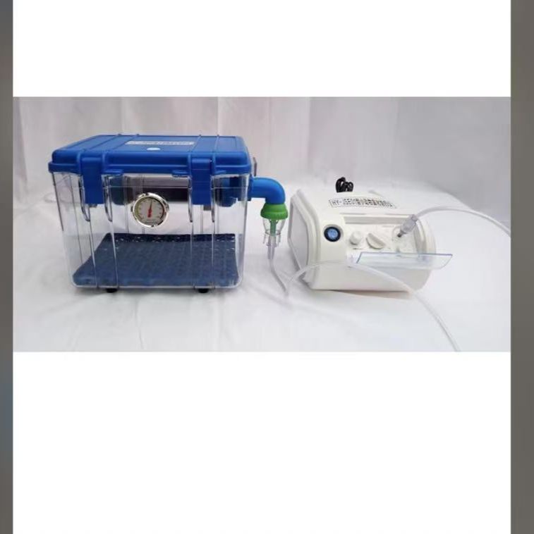
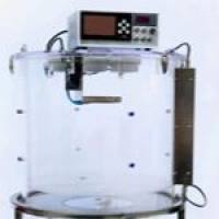
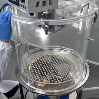
②动物给药舱尺寸可定制:
提供多种规格的独立密闭给药舱,适配小鼠、大鼠、豚鼠等不同体型的实验动物。透明材质便于实时观察,并实现多只动物并行给药,确保实验条件的一致性,极大提升科研效率。
③操作简易,安全高效
可精确设定雾化时间、给药气流速率等关键参数。系统配备高效过滤器,对排出废气进行净化,有效防止药物气溶胶在实验室环境中扩散,保障科研人员的操作安全。
④ 多个知名学术机构购买,并且已经发表多篇高质量论文
(1)广州中医药大学第一附属医院,COMMUNICATIONS BIOLOGY | (2024) 7:181 |,Bronchial epithelial transcriptomics and experimental validation reveal asthma severity[1]related neutrophilc signatures and potential treatments
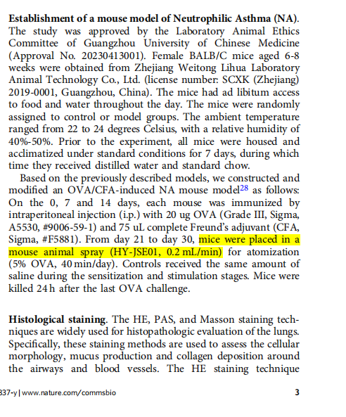
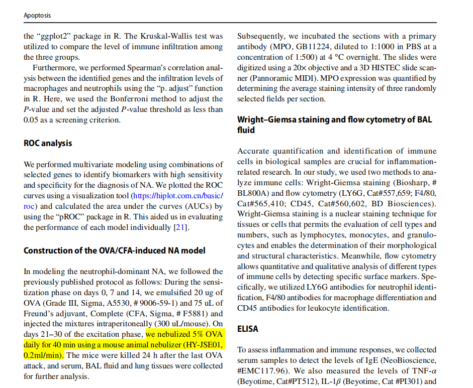
(2)广州中医药大学岭南医学研究中心,Apoptosis,ITGAM‑macrophage modulation as a potential strategy for treating neutrophilic Asthma: insights from bioinformatics analysis and in vivo
experiments
(3)上海交通大学医学院附属新华医院,Dovepress,
Active-Ingredient Screening and Synergistic Action Mechanism of Shegan Mixture for Anti-Asthma Effects Based on Network Pharmacology in a Mouse Model of Asthma
(4)福建医科大学药学院天然药物药理学重点实验室,使用北京元森凯德生物技术有限公司(BEIJING YSKD BIO-TECHNOLOGY CO.,LTD),简称元森凯德(YSKD)研制生产的小动物雾化给药仪HY-JSE01 jet nebulizer (YSKD Biotechnology Co., Ltd., Beijing, China),在中科院1区Top期刊Analytical Chemistry 发表用于新型抗哮喘吸入抗体肺组织分布研究的多功能金纳米簇高分论文。
风险提示:丁香通仅作为第三方平台,为商家信息发布提供平台空间。用户咨询产品时请注意保护个人信息及财产安全,合理判断,谨慎选购商品,商家和用户对交易行为负责。对于医疗器械类产品,请先查证核实企业经营资质和医疗器械产品注册证情况。
 文献和实验
文献和实验DNP. Any unentrapped DNP was removed by filtration through a
0.22 μm filter membrane (Millipore, St. Louis, MO, USA), followed
by gel filtration through Sepharose (CL-4B, Solarbio, China).
GPQ-EL-DNP was prepared using a membrane extrusion method.
L-929 cell-derived exosomes were harvested by ultracentrifugation.23
Briefly, L-929 cells were cultured in exosome-free medium for 48 h,
and the culture medium was centrifuged at 300g for 10 min, 2000g for
20 min, and 10000g for 30 min at 4 °C to remove cells, cell debris,
and microvesicles. Finally, the supernatant was centrifuged at 120000g
for 60 min to obtain a precipitate consisting of exosomes. The pellets
were rinsed and ultracentrifuged, then resuspended in sterile PBS.
The protein concentration of the exosomes was determined by BCA
assay, and the concentration was adjusted to 200 μg/mL. Exosome
markers, CD63 and HSP70, were detected by Western blotting
analysis. A 1 mL solution containing mixed exosomes and GPQ-L-
DNP (protein and lipid weight ratio of 1:5) was sonicated and then
extruded 10 times through a 200 nm polycarbonate membrane (Mini-
Extruder, Avanti Polar Lipoids). The successful synthesis of GPQ-EL-
DNP was further confirmed by FTIR (Nicolet 6700, Thermo Electron
Co., Waltham, MA, USA). Coumarin-6-loaded liposomes, DiD-
loaded liposomes, and SIM-loaded liposomes were prepared using the
same method, except 2% coumarin-6, DiD (ThermoFisher, Waltham,
MA, USA), or 5 mg of SIM was added in chloroform to prepare the
corresponding nanoparticles. The fluorescence intensity of coumarin-
6 and DiD showed a linear relationship with the concentration
(Figure S7A,B). L-DNP/SIM and GPQ-EL-DNP/SIM were prepared
with 3 mg of DNP and 1.5 mg of SIM dissolved in chloroform.
The particle size, polydispersity index, and zeta potential of the
exosomes and liposomes were determined by dynamic light scattering
using a Zetasizer Nano-ZS90 (Malvern Instruments, Malvern, UK),
and the morphology was observed using a transmission electron
microscope (TEM) (JEM-1400, JEOL, Tokyo, Japan). The
encapsulation efficiencies of DNP and SIM in nanoparticles were
quantified using HPLC (LC-20A, Shimadzu, Tokyo, Japan), with
ultraviolet detection at 260 nm for DNP and at 238 nm for SIM. The
mobile phase consisted of acetonitrile and 1% acetate acid (95:5, v/v)
at a flow rate of 1.0 mL/min at room temperature.
The membrane fusion of exosomes and GPQ-liposomes was
verified by FRET. FRET liposomes containing 2% fluorescence-
labeled phospholipids in a lipid layer involved equal amounts of the
electron donor PE-NBD (λex/λem = 488/525 nm) and the electron
acceptor PE-RhB (λex/λem = 560/583 nm). Before and after fusion
with exosomes, the emission spectra of the FRET liposomes were
depicted using a fluorescence spectrometer (F-4600, Hitachi, Tokyo,
Japan) and ranged from 500 to 700 nm at an excitation wavelength of
488 nm. The FRET efficiency was calculated by the following
equation: % FRET efficiency = Fa/(Fa + Fd) × 100%. Fa indicates the
emission fluorescence of RhB, and Fd indicates the emission
fluorescence of NBD.
MMP-9 Responsiveness of GPQ-EL. In vitro release testing of
GPQ-EL was performed using a dialysis membrane diffusion
technique. Coumarin-6-loaded GPQ-EL in medium with or without
MMP-9 (5 nM) was added to a dialysis bag (molecular weight cutoff,
3.5 kDa) and then placed in HEPES (pH 7.4) supplemented with
0.3% sodium dodecyl sulfate maintained at 37 °C with constant
stirring at 100 rpm. At predetermined time intervals, the fluorescence
intensity of coumarin-6 outside the dialysis bag was measured by
fluorescence spectroscopy, and the fresh dissolution medium was
replenished.
The responsiveness of MMP-9 was also verified in L-929 cells with
high MMP-9 expression. L-929 cells (5 × 10⁵/well) were seeded in a
six-well plate containing glass coverslips. After 24 h of incubation,
glucose (22 mM) and homocysteine (100 μM) were added for an
addition 6 h of incubation to stimulate MMP-9 expression, and then
FRET liposomes were added. After incubation at 37 °C for 4 h, the
cells were subjected to a confocal laser scanning microscopy (TCS
SP8, Leica, Wetzlar, Germany) for detection in the NBD (525−550
nm) and RhB (560−600 nm) channels at an excitation wavelength of
488 and 560 nm, respectively.
增加了药物吸收的有效面积。由于粘膜细胞下有着丰富的血管和淋巴管,药物通过粘膜吸收后可直接进入体循环。此外,鼻腔内酶的代谢作用远远小于胃肠道,因此,鼻腔给药系统正日 益受到人们的重视,包括肽类和蛋白质类药物的研究。在众多给药剂型中,喷雾剂是比较常见的剂型,仅通过雾化装置借助压缩空气产生的动力使药液雾化并喷出。由于喷雾剂不含抛射剂,无需使用耐压容器,目前应用领域越来越广泛。在鼻喷剂研究过程中,对于鼻喷剂粒度分布大小有两个因素影响至关重要,即药物配方和喷射装置。您通过以下一些模拟实验可了解到激光
注射 0.2ml2 激发模型组 于实验第 15 天 将大鼠置于 20cm×20cm×15cm 大小的密闭容器中由 PARI BOY高频雾化器提供雾化动力 以 1%OVA GradeII 进行雾化吸入激发 每次雾化 30min 连续 6 天.3 观察指标和检测方法3. 1 外周血单个核细胞(PBMC)的制备及培养大鼠无菌心脏穿刺采血 2ml 肝素抗凝 吸取血浆 100ìl于-20o冻存待检 IgE剩余部分与等量 PBS 液充分混匀 在一试管中加入适量淋巴细胞分离液 Ficoll用滴管将血液沿管壁缓慢叠加
入的菌液易漏,后来尝试用50ul的加样器每次取20ul,采取多次注射的办法。做此类实验主要是注意感染的药物或液体不要漏出来,可以采取多次注射的办法。八、大鼠鼻腔给药1.有滴鼻和喷雾两种常见方式喷雾其实就是雾化吸入。滴鼻给药没有办法达到雾化吸入的效果。雾化吸入需要有雾化设备,一般医院的都有,但是医院的如果借不出来,自己家里的加湿器也可以凑合。雾化给药的时候,要把大鼠放在一个相对比较密闭的的容器中(当然要有透气孔),让大鼠尽可能多地接触药物,但是好象没有专门的这种容器,一般都是自制的,材料最好是有机
 技术资料
技术资料暂无技术资料 索取技术资料

