


- 询价
- YSKD
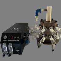
- 大鼠气溶胶给药系统
- 2026年04月22日

企业认证

万千商家帮你免费找货
0 人在求购买到急需产品
- 详细信息
- 询价记录
- 文献和实验
- 技术资料
- 保修期:
12个月
- 现货状态:
现货
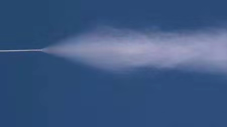
性能特点:
精确定量
较气管内滴入在各肺叶中分布更均匀
直达肺部、易于操作
更安全的提供高浓度
可输送液体、干粉样品
风险提示:丁香通仅作为第三方平台,为商家信息发布提供平台空间。用户咨询产品时请注意保护个人信息及财产安全,合理判断,谨慎选购商品,商家和用户对交易行为负责。对于医疗器械类产品,请先查证核实企业经营资质和医疗器械产品注册证情况。
- 作者
- 内容
- 询问日期
 文献和实验
文献和实验Treating Multiorgan Fibrosis
Qiang Long, Zehua Liu, Qianwen Shao, Hongpeng Shi, Shixing Huang, Chenyu Jiang,
Bei Qian, Yiming Zhong, Xiaojun He, Xiaogang Xiang, Yang Yang, Bing Li, Xiaoxiang Yan,
Qiang Zhao,* Xiaoli Wei,* Hélder A. Santos,* and Xiaofeng Ye*
Fibrotic diseases remain a substantial health burden with few therapeutic
approaches. A hallmark of fibrosis is the aberrant activation and accumulation
of myofibroblasts, which is caused by excessive profibrotic cytokines.
Conventional anticytokine therapies fail to undergo clinical trials, as simply
blocking a single or several antifibrotic cytokines cannot abrogate the
profibrotic microenvironment. Here, biomimetic nanoparticles based on
autologous skin fibroblasts are customized as decoys to neutralize multiple
fibroblast-targeted cytokines. By fusing the skin fibroblast membrane onto
poly(lactic-co-glycolic) acid cores, these nanoparticles, termed fibroblast
membrane-camouflaged nanoparticles (FNPs), are shown to effectively
scavenge various profibrotic cytokines, including transforming growth
factor-휷, interleukin (IL)-11, IL-13, and IL-17, thereby modulating the
profibrotic microenvironment. FNPs are sequentially prepared into multiple
formulations for different administration routines. As a proof-of-concept, in
three independent animal models with various organ fibrosis (lung fibrosis,
liver fibrosis, and heart fibrosis), FNPs effectively reduce the accumulation of
myofibroblasts, and the formation of fibrotic tissue, concomitantly restoring
organ function and indicating that FNPs are a potential broad-spectrum
therapy for fibrosis management.
Q. Long, H. Shi, S. Huang, C. Jiang, B. Qian, Y. Zhong, X. He, Q. Zhao,
X. Ye
Department of Cardiovascular Surgery
Ruijin Hospital
Shanghai Jiao Tong University School of Medicine
Shanghai 200025, China
E-mail: zq11607@rjh.com.cn; yxf11612@rjh.com.cn
Z. Liu, H. A. Santos
Department of Biomedical Engineering, W.J. Kolff Institute for
Biomedical Engineering and Materials Science
University Medical Center Groningen/University of Groningen
Ant. Deusinglaan 1, Groningen 9713 AV, The Netherlands
E-mail: h.a.santos@umcg.nl
The ORCID identification number(s) for the author(s) of this article
can be found under https://doi.org/10.1002/advs.202200856
© 2022 The Authors. Advanced Science published by Wiley-VCH GmbH.
This is an open access article under the terms of the Creative Commons
Attribution License, which permits use, distribution and reproduction in
any medium, provided the original work is properly cited.
DOI: 10.1002/advs.202200856
1. Introduction
Fibrosis, or disordered fibrotic tissue formation, is characterized by the abnormal
fibroblast activation that induces excessive extracellular matrix (ECM) remodeling
and primarily accounts for multiple organ
dysfunctions.[1] The pervasive occurrence
of fibrosis in almost all diseases generates
a large healthcare burden worldwide. However, the clinical benefits of antifibrotic therapy through small molecules, such as pirfenidone and nintedanib, are usually offset
by their modest therapeutic efficacy, limited
indications and severe side effects.[2] Therefore, alternative clinical intervention modalities to target fibrosis are urgently needed.
Considering the central role of myofibroblast activation and proliferation in
fibrosis establishment,[3] recent breakthroughs have focused on the ablation
of progressive myofibroblast activation
through autologous cell-based therapy.
For example, autologous chimeric antigen
Z. Liu, H. A. Santos
Drug Research Program
Division of Pharmaceutical Chemistry and Technology
Faculty of Pharmacy
University of Helsinki
Helsinki FI-00014, Finland
Q. Shao, X. Wei
Department of Pharmacology
School of Basic Medical Sciences
Fudan University
Shanghai 200032, China
E-mail: xlwei@fudan.edu.cn
X. Xiang
Department of Infectious Diseases
Ruijin Hospital
Shanghai Jiao Tong University School of Medicine
Shanghai 200025, China
Y. Yang
Department of Thoracic Surgery
Shanghai Pulmonary Hospital
School of Medicine
Tongji University
Shanghai 200000, China
Adv. Sci. 2022, 9, 2200856 2200856 (1 of 14) © 2022 The Authors. Advanced Science published by Wiley-VCH GmbH
性呼吸,直到呼吸停止而死亡。如果动物致敏程度较轻或诱发时鸡蛋白喷雾的浓度很快,则只发生一时性的支气管痉挛,并不死亡。如改用组织胺喷雾,则不必予先致敏,就能引起豚鼠支气管痉挛。组织胺用量依雾室大小而定,在83~103容量时,1∶1000组织胺的用量为0.5~1ml。狗每周两次暴露于犬弓蛔虫(Toxocaracanis)、猪蛔虫(Ascaris suum)或混合草籽浸出物的气溶胶中可引起实验性哮喘。给用10-8稀释猪蛔虫浸出物皮试阳性狗以猪蛔虫气溶胶吸入,也可引起哮喘。(五)实验性矽肺模型常选用大鼠、家兔或狗、猴
一、慢性支气管肺炎模型 常选用大鼠、豚鼠或猴吸入刺激性气体(如二氧化硫、氯、氨水、烟雾等)复制人类慢性气管炎。现发现猪粘膜下腺体与人类相似,且经常发生气管炎及肺炎,故认为是复制人类慢性气管炎较合适的动物。用去甲肾上腺素可以引起与人类相似的气管腺体肥大。 二、肺气肿模型 给兔等动物气管内或静脉内注射一定量木瓜蛋白酶、菠罗蛋白酶(Bromelin)、败血酶(Alcalas)、胰蛋白酶(Trypsin)、致热溶解酶(Thermolysin),以及由脓性痰和白细胞分离出来的蛋白
在免疫学研究中,常选用以下的几种实验动物:(一)大鼠:大鼠对绵羊红细胞和牛r球蛋白的免疫反应有品系差异。大鼠有抗体IgE,蠕虫感染常能诱发大量的IgE抗体,它们存在于血液循环中,常规的免疫法只能使大鼠产生少量的抗体,在体内存在的时间较短。百日咳杆菌免疫大鼠主要产生IgE,如在此抗原中加入福氏完全佐剂,免疫大鼠则产生IgGa。(二)小鼠:小鼠的免疫球蛋白有IgM、IgA、IgE、IgG1、IgG2a和IgG2b。小鼠很少见到典型的迟发型变态反应,也不象其它动物那样有规律。能诱发速发型变态
 技术资料
技术资料暂无技术资料 索取技术资料

