

- ¥11452.50
- Lifeline

- 美国
- FC-0017
- 2026年03月01日

企业认证
相关产品推荐更多 >

万千商家帮你免费找货
0 人在求购买到急需产品
- 详细信息
- 文献和实验
- 技术资料
- 英文名:
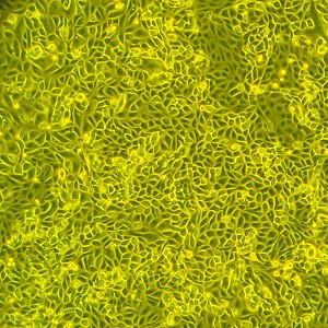
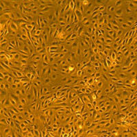
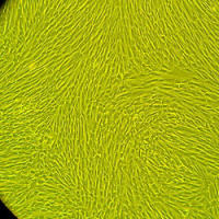
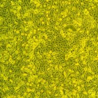
Normal Human Renal Mixed Epithelial Cells, Primary
- 细胞类型:
人正常原代细胞
- 物种来源:
人源
- 器官来源:
人源
- 运输方式:
干冰或液氮
- 年限:
液氮储存10年以上
- 生长状态:
冻存
- 规格:
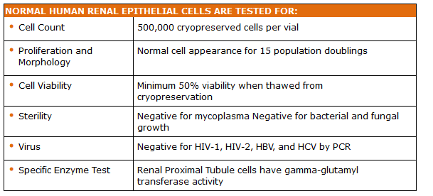
500,000 cells/vial
Lifeline® normal Human Renal Mixed Epithelial Cells, when grown in Lifeline RenaLife™ Medium, provide an ideal low-serum culture model for the study of renal function, fibrosis, inflammation, metabolism, nephrotoxicity or cancer. Additionally, our normal human renal cells can be used for drug development or screening.
Lifeline Renal Mixed Epithelial Cells are cryopreserved as primary cells to ensure the highest viability and plating efficiency.
- Primary cells have been isolated from human kidney tissue, plated onto culture vessels, expanded once, harvested and cryopreserved.
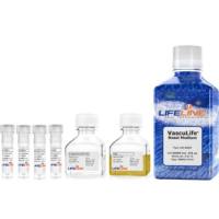
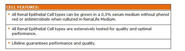
Our Renal Epithelial Cells are quality tested in RenaLife™ Medium to ensure optimal reduced-serum growth over a period of at least 15 population doublings at rates equal to or greater than serum-supplemented medium.
- Renal Mixed Epithelial Cells can be grown in a 0.5% serum medium without phenol red or antimicrobials when cultured in RenaLife™ Medium.
Lifeline® Renal Mixed Epithelial Cells are not exposed to antimicrobials or phenol red when cultured in RenaLife™ Medium, an advantage since these supplements can cause cell stress and “masking effects” that may negatively impact experimental results. Lifeline® offers these traditional supplements; however, they are not needed, or recommended, to achieve optimal cell performance in most research applications.
500,000 cells per vial
Quality Testing for Guaranteed Consistency and Reproducible Results
Lifeline® Cell Technology manufactures products using the highest quality raw materials and incorporates extensive quality assurance in every production run. Exacting standards and production procedures ensure consistent performance.
风险提示:丁香通仅作为第三方平台,为商家信息发布提供平台空间。用户咨询产品时请注意保护个人信息及财产安全,合理判断,谨慎选购商品,商家和用户对交易行为负责。对于医疗器械类产品,请先查证核实企业经营资质和医疗器械产品注册证情况。
 文献和实验
文献和实验的分枝尖端,形成帽状间充质,促进帽状间充质中的肾祖细胞增殖分化,形成聚集(Pre-tubular aggregate,PA)。随后,PA 和 UB 分别进行间充质-上皮细胞转化(Mesenchymal-epithelial transition, MET)和小管生成,进而形成肾单位【2】。目前广泛用于肾脏研究的人肾脏细胞系忽略了细胞外微环境,与在体的器官类型和整体特性仍存在较大差异,不能充分体现出肾脏的作用及其特点,而三维培养的肾类器官模型为肾脏的研究提供了新途径。人源多能干细胞衍生的肾脏类器官
应该是肾小球内(脏)层上皮细胞,这株细胞目前只有私人收藏,全球细胞库没有严格意义上的足细胞可供出售。 在文献中提到了“人肾小球系膜细胞系(human mesangial cell line,HMCL)由英国伦敦皇家公立医院肾脏中心阮雄中博士惠赠-----HMCL 为用T2SV40 及H2ras 原癌基因转染的永生系,保持正常人肾系膜细胞基本的形态学和生物学特征"。 因为足细胞很难找到,国内一些研究者已改用肾小管上皮细胞hk-2
激光捕获显微切割技术与生物芯片的联合应用-提高检验组织样品的特异性
人体组织由细胞和细胞间基质组成,肿瘤也不例外。除了肿瘤细胞之外,肿瘤还含有血管、结缔组织等基质。目前,常规对肿瘤样品基因表达量的研究实际上是对肿瘤细胞和基质混合物的研究。基质细胞往往没有癌变,因此对肿瘤细胞和基质混合物进行研究往往会掩盖掉肿瘤细胞中并不是很明显的基因表达异常。 最近复旦大学吴莹教授等利用激光捕获显微切割技术(Laser capture microdissection,LCM)很好的解决了这一难题。相关成果发表在2010年第11期的BMC Genomics上。激光捕获显微
 技术资料
技术资料暂无技术资料 索取技术资料

