大家都在搜

手机验证

询价列表

暂时没有已询价产品

SuperSignal ECL
Western Blotting Substrate

企业认证
现货
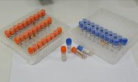
SuperSignal ECL Western Blotting Substrate
12 months
华安生物
4°C
100 mL/200 mL/500 mL
| 规格: | 100 mL | 产品价格: | ¥600.0 |
|---|---|---|---|
| 规格: | 200 mL | 产品价格: | ¥1100.0 |
| 规格: | 500 mL | 产品价格: | ¥2200.0 |
.jpg)
风险提示:丁香通仅作为第三方平台,为商家信息发布提供平台空间。用户咨询产品时请注意保护个人信息及财产安全,合理判断,谨慎选购商品,商家和用户对交易行为负责。对于医疗器械类产品,请先查证核实企业经营资质和医疗器械产品注册证情况。
 询价记录
询价记录