


- ¥880 - 2480
- gelatins
- jlcR1839
- 国内
- 2025年07月15日
- WB,ELISA等
- 人/动物/植物

企业认证
相关产品推荐更多 >


万千商家帮你免费找货
0 人在求购买到急需产品
- 详细信息
- 询价记录
- 文献和实验
- 技术资料
- 供应商:
江西江蓝纯生物试剂有限公司
- 库存:
108
- 克隆性:
单克隆
- 保质期:
1年
- 抗体英文名:
lamin A + C
- 抗体名:
核纤层蛋白A抗体
- 适应物种:
人/动物/植物
- 应用范围:
WB,ELISA等
- 浓度:
1mg/ml
- 保存条件:
-20 °
- 规格:
50ul/100ul/200ul
| 规格: | 50ul | 产品价格: | ¥880.0 |
|---|---|---|---|
| 规格: | 100ul | 产品价格: | ¥1580.0 |
| 规格: | 200ul | 产品价格: | ¥2480.0 |
英文名称 : lamin A + C
中文名称 : 核纤层蛋白A抗体
别 名 : lamin A/C; LMN1; 70 kDa lamin; CDCD1; CDDC; CMD1A; CMT2B1; EMD2; FPL; FPLD; HGPS; IDC; LAMIN A; lamin A/C; LAMIN C; LDP1; LFP; LGMD1B; LMN 1; LMN A; LMN C; LMNA; LMNC; NY REN 32 antigen; PRO1; LMNA_HUMAN; Prelamin-A/C; Renal carcinoma antigen NY-REN-32.
研究领域 : 细胞生物 细胞凋亡
抗体来源 : Rabbit
克隆类型 : Polyclonal
交叉反应 : Human, Mouse, Rat,
产品应用 : WB=1:500-2000 ELISA=1:500-1000 IHC-P=1:400-800 IHC-F=1:400-800 IF=1:100-500 (石蜡切片需做抗原修复)
not yet tested in other applications.
optimal dilutions/concentrations should be determined by the end user.
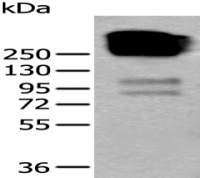
分 子 量 : 73kDa
细胞定位 : 细胞核
性 状 : Lyophilized or Liquid
浓 度 : 1mg/ml
免 疫 原 : KLH conjugated synthetic peptide derived from human lamin A:1-100/664
亚 型 : IgG
纯化方法 : affinity purified by Protein A
储 存 液 : 0.01M TBS(pH7.4) with 1% BSA, 0.03% Proclin300 and 50% Glycerol.
保存条件 : Store at -20 °C for one year. Avoid repeated freeze/thaw cycles. The lyophilized antibody is stable at room temperature for at least one month and for greater than a year when kept at -20°C. When reconstituted in sterile pH 7.4 0.01M PBS or diluent of antibody the antibody is stable for at least two weeks at 2-4 °C.
PubMed : PubMed
产品介绍 : Nuclear lamins form a network of intermediate-type filaments at the nucleoplasmic site of the nuclear membrane. Two main subtypes of nuclear lamins can be distinguished, i.e. A type lamins and B type lamins. The A type lamins comprise a set of three proteins arising from the same gene by alternative splicing, i.e. lamin A, lamin C and lamin Adel 10, while the B type lamins include two proteins arising from two distinct genes, i.e. lamin B1 and lamin B2. Recent evidence has revealed that mutations in A-type lamins give rise to a range of rare but dominant genetic disorders, including Emery-Dreifuss muscular dystrophy, dilated cardiomyopathy with conduction-system disease and Dunnigan-type familial partial lipodystrophy. In addition, the expression of A type lamins coincides with cell differentiation and as A type lamins specifically interact with chromatin, a role in the regulation of differential gene expression has been suggested for A type lamins.
Function:
Lamins are components of the nuclear lamina, a fibrous layer on the nucleoplasmic side of the inner nuclear membrane, which is thought to provide a framework for the nuclear envelope and may also interact with chromatin. Lamin A and C are present in equal amounts in the lamina of mammals. Play an important role in nuclear assembly, chromatin organization, nuclear membrane and telomere dynamics.
Prelamin-A/C can accelerate smooth muscle cell senescence. It acts to disrupt mitosis and induce DNA damage in vascular smooth muscle cells (VSMCs), leading to mitotic failure, genomic instability, and premature senescence
Subunit:
Homodimer of lamin A and lamin C. Interacts with lamin-associated polypeptides IA, IB and TMPO-alpha, RB1 and with emerin. Interacts with SREBF1, SREBF2, SUN2 and TMEM43. Proteolytically processed isoform A interacts with NARF. Interacts with SUN1. Prelamin-A/C interacts with EMD. Interacts with MLIP; may regulate MLIP localization to the nucleus envelope. Interacts with DMPK; may regulate nuclear envelope stability.
Subcellular Location:
Nucleus. Nucleus envelope. Note=Farnesylation of prelamin-A/C facilitates nuclear envelope targeting and subsequent cleaveage by ZMPSTE24/FACE1 to remove the farnesyl group produces mature lamin-A/C, which can then be inserted into the nuclear lamina. EMD is required for proper localization of non-farnesylated prelamin-A/C.
Tissue Specificity:
In the arteries, prelamin-A/C accumulation is not observed in young healthy vessels but is prevalent in medial vascular smooth muscle cells (VSMCs) from aged individuals and in atherosclerotic lesions, where it often colocalizes with senescent and degenerate VSMCs. Prelamin-A/C expression increases with age and disease. In normal aging, the accumulation of prelamin-A/C is caused in part by the down-regulation of ZMPSTE24/FACE1 in response to oxidative stress.
Post-translational modifications:
Increased phosphorylation of the lamins occurs before envelope disintegration and probably plays a role in regulating lamin associations.
Proteolytic cleavage of the C-terminal of 18 residues of prelamin-A/C results in the production of lamin-A/C. The prelamin-A/C maturation pathway includes farnesylation of CAAX motif, ZMPSTE24/FACE1 mediated cleavage of the last three amino acids, methylation of the C-terminal cysteine and endoproteolytic removal of the last 15 C-terminal amino acids. Proteolytic cleavage requires prior farnesylation and methylation, and absence of these blocks cleavage.
Sumoylation is necessary for the localization to the nuclear envelope.
Farnesylation of prelamin-A/C facilitates nuclear envelope targeting.
DISEASE:
Defects in LMNA are the cause of Emery-Dreifuss muscular dystrophy type 2, autosomal dominant (EDMD2) [MIM:181350]. A degenerative myopathy characterized by weakness and atrophy of muscle without involvement of the nervous system, early contractures of the elbows, Achilles tendons and spine, and cardiomyopathy associated with cardiac conduction defects.
Defects in LMNA are the cause of Emery-Dreifuss muscular dystrophy type 3, autosomal recessive (EDMD3) [MIM:181350].
Defects in LMNA are the cause of cardiomyopathy dilated type 1A (CMD1A) [MIM:115200]. Dilated cardiomyopathy is a disorder characterized by ventricular dilation and impaired systolic function, resulting in congestive heart failure and arrhythmia. Patients are at risk of premature death.
Defects in LMNA are the cause of familial partial lipodystrophy type 2 (FPLD2) [MIM:151660]; also known as familial partial lipodystrophy Dunnigan type. A disorder characterized by the loss of subcutaneous adipose tissue in the lower parts of the body (limbs, buttocks, trunk). It is accompanied by an accumulation of adipose tissue in the face and neck causing a double chin, fat neck, or cushingoid appearance. Adipose tissue may also accumulate in the axillae, back, labia majora, and intraabdominal region. Affected patients are insulin-resistant and may develop glucose intolerance and diabetes mellitus after age 20 years, hypertriglyceridemia, and low levels of high density lipoprotein cholesterol.
Defects in LMNA are the cause of limb-girdle muscular dystrophy type 1B (LGMD1B) [MIM:159001]. LGMD1B is an autosomal dominant degenerative myopathy with age-related atrioventricular cardiac conduction disturbances, dilated cardiomyopathy, and the absence of early contractures. LGMD1B is characterized by slowly progressive skeletal muscle weakness of the hip and shoulder girdles. Muscle biopsy shows mild dystrophic changes.
Defects in LMNA are the cause of Charcot-Marie-Tooth disease type 2B1 (CMT2B1) [MIM:605588]. CMT2B1 is a form of Charcot-Marie-Tooth disease, the most common inherited disorder of the peripheral nervous system. Charcot-Marie-Tooth disease is classified in two main groups on the basis of electrophysiologic properties and histopathology: primary peripheral demyelinating neuropathy or CMT1, and primary peripheral axonal neuropathy or CMT2. Neuropathies of the CMT2 group are characterized by signs of axonal regeneration in the absence of obvious myelin alterations, normal or slightly reduced nerve conduction velocities, and progressive distal muscle weakness and atrophy. CMT2B1 inheritance is autosomal recessive.
Defects in LMNA are the cause of Hutchinson-Gilford progeria syndrome (HGPS) [MIM:176670]. HGPS is a rare genetic disorder characterized by features reminiscent of marked premature aging. Note=HGPS is caused by the toxic accumulation of a mutant form of lamin-A/C. This mutant protein, called progerin, acts to deregulate mitosis and DNA damage signaling, leading to premature cell death and senescence. Progerin lacks the conserved ZMPSTE24/FACE1 cleavage site and therefore remains permanently farnesylated. Thus, although it can enter the nucleus and associate with the nuclear envelope, it cannot incorporate normally into the nuclear lamina.
Defects in LMNA are the cause of cardiomyopathy dilated with hypergonadotropic hypogonadism (CMDHH) [MIM:212112]. A disorder characterized by the association of genital anomalies, hypergonadotropic hypogonadism and dilated cardiomyopathy. Patients can present other variable clinical manifestations including mental retardation, skeletal anomalies, scleroderma-like skin, graying and thinning of hair, osteoporosis. Dilated cardiomyopathy is characterized by ventricular dilation and impaired systolic function, resulting in congestive heart failure and arrhythmia.
Defects in LMNA are the cause of mandibuloacral dysplasia with type A lipodystrophy (MADA) [MIM:248370]. A disorder characterized by mandibular and clavicular hypoplasia, acroosteolysis, delayed closure of the cranial suture, progeroide appearance, partial alopecia, soft tissue calcinosis, joint contractures, and partial lipodystrophy with loss of subcutaneous fat from the extremities. Adipose tissue in the face, neck and trunk is normal or increased.
Defects in LMNA are a cause of lethal tight skin contracture syndrome (LTSCS) [MIM:275210]; also known as restrictive dermopathy (RD). Lethal tight skin contracture syndrome is a rare disorder mainly characterized by intrauterine growth retardation, tight and rigid skin with erosions, prominent superficial vasculature and epidermal hyperkeratosis, facial features (small mouth, small pinched nose and micrognathia), sparse/absent eyelashes and eyebrows, mineralization defects of the skull, thin dysplastic clavicles, pulmonary hypoplasia, multiple joint contractures and an early neonatal lethal course. Liveborn children usually die within the first week of life. The overall prevalence of consanguineous cases suggested an autosomal recessive inheritance.
Defects in LMNA are the cause of heart-hand syndrome Slovenian type (HHS-Slovenian) [MIM:610140]. Heart-hand syndrome (HHS) is a clinically and genetically heterogeneous disorder characterized by the co-occurrence of a congenital cardiac disease and limb malformations.
Defects in LMNA are the cause of muscular dystrophy congenital LMNA-related (MDCL) [MIM:613205]. It is a form of congenital muscular dystrophy. Patients present at birth, or within the first few months of life, with hypotonia, muscle weakness and often with joint contractures.
Similarity:
Belongs to the intermediate filament family.
SWISS:
P02545
Gene ID:
4000
Important Note:
This product as supplied is intended for research use only, not for use in human, therapeutic or diagnostic applications.
核膜标志物(Nuclear Envelope Marker)
核纤层蛋白(lamin) 是紧贴核内膜的一层厚度为20~50nm的纤维蛋白层或纤维网络。核纤层与细胞质骨架、核骨架连成一个整体,一般认为核纤层将核被膜和染色质提供了结构支架。有学者研究认为:lamin蛋白与细胞凋亡及衰老有关联,它包括:核纤层蛋白A、核纤层蛋白B、核纤层蛋白C几个不同亚型的蛋白。
风险提示:丁香通仅作为第三方平台,为商家信息发布提供平台空间。用户咨询产品时请注意保护个人信息及财产安全,合理判断,谨慎选购商品,商家和用户对交易行为负责。对于医疗器械类产品,请先查证核实企业经营资质和医疗器械产品注册证情况。
- 作者
- 内容
- 询问日期
 文献和实验
文献和实验脊椎动物细胞中有三种类型的核纤层蛋白(A,B,C), 核纤层蛋白A和C是由同一个转录单位编码的, 只不过是通过可变剪接形成不同的mRNA。它们在肽链上的差别是:核纤层蛋白A的C末端比核纤层蛋白C的C末端多133个氨基酸残基。核纤层蛋白B是由另一个转录单位编码的, 通过转录后的修饰,在羧基端添加了疏水的异丙基, 添加的脂肪酸帮助核纤层蛋白B插入到核膜的内脂层。三种类型的核纤层蛋白都以二聚体的形式存在, 有球形的头和尾部结构域以及一个杆状的α螺旋中心。这些核纤层蛋白二聚体以头-头、尾-尾相接
鸭免疫球蛋白A抗体(IgA-Ab)酶联免疫分析(ELISA)
鸭 免疫球蛋白A 抗体 ( IgA-Ab ) 酶联免疫 分析(ELISA) 试剂 盒使用说明书 本试剂仅供研究使用 目的:本试剂盒用于测定鸭血清,血浆及相关液体样本中 免疫球蛋白A抗体(IgA-Ab) 的 含量。 实验原理 : 本试剂盒应用双抗 原 夹心法测定 标本 中 鸭 免疫球蛋白 A抗体(IgA-Ab) 水平。用纯化的 免疫球蛋白 A抗体(IgA-Ab) 抗 原 包被微孔板,制成固相抗 原 ,往包被 抗原 的微孔中依次
相关专题 免疫胶体金技术专题 摘 要: 目的 建立一种快速、简易的检测血清中抗幽门螺杆菌(Hp)细胞毒素相关蛋白A(CagA)抗体的胶体金免疫层析法(GICA)。 方法 采用柠檬酸三钠还原法制备胶体金颗粒,标记葡萄球菌A蛋白(SPA),将重组 的CagA抗原划线固定于硝酸纤维素膜上,制成免疫层析检测试条。 血清中IgG与测试条上金标记物结合后沿着硝酸纤维素膜移动,与膜上的固相抗体结合形成肉眼可见的红色线条。 结果 用GICA

