


- ¥880 - 2480
- gelatins
- jlcR0076
- 国内
- 2026年03月29日
- WB,ELISA等
- 人/动物/植物

企业认证
相关产品推荐更多 >


万千商家帮你免费找货
0 人在求购买到急需产品
- 详细信息
- 文献和实验
- 技术资料
- 供应商:
江西江蓝纯生物试剂有限公司
- 库存:
38
- 克隆性:
单克隆
- 保质期:
1年
- 抗体英文名:
beta-Amyloid 1-42(CT)
- 抗体名:
β淀粉样肽/Aβ42抗体
- 适应物种:
人/动物/植物
- 应用范围:
WB,ELISA等
- 浓度:
1mg/ml
- 保存条件:
-20 °
- 规格:
50ul/100ul/200ul
| 规格: | 50ul | 产品价格: | ¥880.0 |
|---|---|---|---|
| 规格: | 100ul | 产品价格: | ¥1580.0 |
| 规格: | 200ul | 产品价格: | ¥2480.0 |
英文名称 : beta-Amyloid 1-42(CT)
中文名称 : β淀粉样肽/Aβ42抗体
别 名 : beta Amyloid(1-42); beta Amyloid 1-42; beta-Amyloid 1-42; Amyloid 1-42; beta-Amyloid 42; A4; AAA; ABETA; ABPP; AD1; Alzheimers Disease Amyloid Protein; Amyloid B; Amyloid Beta A4 Protein Precursor; Amyloid Beta; Amyloid of Aging and Alzheimer Disease; APP; APPI; B Amyloid; Beta APP; Cerebral Vascular Amyloid Peptide; CTFgamma; CVAP; PN II; PN2; PreA4; Protease nexin II; A beta; A4_HUMAN.
研究领域 : 细胞生物 免疫学 神经生物学
抗体来源 : Rabbit
克隆类型 : Polyclonal
交叉反应 : Human, Mouse, Rat, Dog, Pig, Cow, Horse, Rabbit,
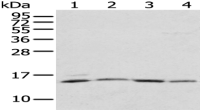
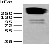
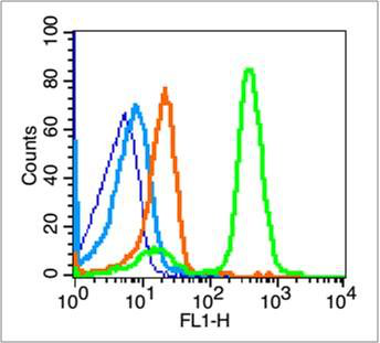
产品应用 : ELISA=1:500-1000 IHC-P=1:400-800 IHC-F=1:400-800 Flow-Cyt=0.2μg/Test IF=1:100-500 (石蜡切片需做抗原修复)
not yet tested in other applications.
optimal dilutions/concentrations should be determined by the end user.
分 子 量 : 4.3/75kDa
细胞定位 : 细胞膜
性 状 : Lyophilized or Liquid
浓 度 : 1mg/ml
免 疫 原 : KLH conjugated synthetic peptide derived from human beta-Amyloid:35-42/42 <Cytoplasmic>
亚 型 : IgG
纯化方法 : affinity purified by Protein A
储 存 液 : 0.01M TBS(pH7.4) with 1% BSA, 0.03% Proclin300 and 50% Glycerol.
保存条件 : Store at -20 °C for one year. Avoid repeated freeze/thaw cycles. The lyophilized antibody is stable at room temperature for at least one month and for greater than a year when kept at -20°C. When reconstituted in sterile pH 7.4 0.01M PBS or diluent of antibody the antibody is stable for at least two weeks at 2-4 °C.
PubMed : PubMed
产品介绍 : The cerebral and vascular plaques associated with Alzheimer's disease are mainly composed of Amyloid beta peptides. beta Amyloid is derived from cleavage of the Amyloid precursor protein and varies in length from 39 to 43 amino acids. beta Amyloid [1-40], beta Amyloid [1-42], and beta Amyloid [1-43] peptides result from cleavage of Amyloid precursor protein after residues 40, 42, and 43, respectively. The cleavage takes place by gamma-secretase during the last Amyloid precursor protein processing step. beta Amyloid [1-40], beta Amyloid [1-42], and beta Amyloid [1-43] peptides are major constituents of the plaques and tangles that occur in Alzheimer's disease. beta Amyloid antibodies and peptides have been developed as tools for elucidating the biology of Alzheimer's disease.
Function:
Functions as a cell surface receptor and performs physiological functions on the surface of neurons relevant to neurite growth, neuronal adhesion and axonogenesis. Involved in cell mobility and transcription regulation through protein-protein interactions. Can promote transcription activation through binding to APBB1-KAT5 and inhibits Notch signaling through interaction with Numb. Couples to apoptosis-inducing pathways such as those mediated by G(O) and JIP. Inhibits G(o) alpha ATPase activity. Acts as a kinesin I membrane receptor, mediating the axonal transport of beta-secretase and presenilin 1. Involved in copper homeostasis/oxidative stress through copper ion reduction. In vitro, copper-metallated APP induces neuronal death directly or is potentiated through Cu(2+)-mediated low-density lipoprotein oxidation. Can regulate neurite outgrowth through binding to components of the extracellular matrix such as heparin and collagen I and IV. The splice isoforms that contain the BPTI domain possess protease inhibitor activity. Induces a AGER-dependent pathway that involves activation of p38 MAPK, resulting in internalization of amyloid-beta peptide and leading to mitochondrial dysfunction in cultured cortical neurons.
Beta-amyloid peptides are lipophilic metal chelators with metal-reducing activity. Bind transient metals such as copper, zinc and iron. In vitro, can reduce Cu(2+) and Fe(3+) to Cu(+) and Fe(2+), respectively. Beta-amyloid 42 is a more effective reductant than beta-amyloid 40. Beta-amyloid peptides bind to lipoproteins and apolipoproteins E and J in the CSF and to HDL particles in plasma, inhibiting metal-catalyzed oxidation of lipoproteins. Beta-APP42 may activate mononuclear phagocytes in the brain and elicit inflammatory responses. Promotes both tau aggregation and TPK II-mediated phosphorylation. Interaction with overexpressed HADH2 leads to oxidative stress and neurotoxicity.
Appicans elicit adhesion of neural cells to the extracellular matrix and may regulate neurite outgrowth in the brai.
The gamma-CTF peptides as well as the caspase-cleaved peptides, including C31, are potent enhancers of neuronal apoptosis.
N-APP binds TNFRSF21 triggering caspase activation and degeneration of both neuronal cell bodies (via caspase-3) and axons (via caspase-6).
Subunit:
Binds, via its C-terminus, to the PID domain of several cytoplasmic proteins, including APBB family members, the APBA family, MAPK8IP1, SHC1 and, NUMB and DAB. Binding to DAB1 inhibits its serine phosphorylation (By similarity). Also interacts with GPCR-like protein BPP, FPRL1, APPBP1, IB1, KNS2 (via its TPR domains), APPBP2 (via BaSS) and DDB1. In vitro, it binds MAPT via the MT-binding domains. Associates with microtubules in the presence of ATP and in a kinesin-dependent manner. Interacts, through a C-terminal domain, with GNAO1. Amyloid beta-42 binds CHRNA7 in hippocampal neurons. Beta-amyloid associates with HADH2. Soluble APP binds, via its N-terminal head, to FBLN1. Interacts with CPEB1 and AGER. Interacts with ANKS1B and TNFRSF21. Interacts with ITM2B. Interacts with ITM2C. Interacts with IDE. Can form homodimers; this is promoted by heparin binding.
Subcellular Location:
Membrane; Single-pass type I membrane protein. Membrane, clathrin-coated pit. Note=Cell surface protein that rapidly becomes internalized via clathrin-coated pits. During maturation, the immature APP (N-glycosylated in the endoplasmic reticulum) moves to the Golgi complex where complete maturation occurs (O-glycosylated and sulfated). After alpha-secretase cleavage, soluble APP is released into the extracellular space and the C-terminal is internalized to endosomes and lysosomes. Some APP accumulates in secretory transport vesicles leaving the late Golgi compartment and returns to the cell surface. Gamma-CTF(59) peptide is located to both the cytoplasm and nuclei of neurons. It can be translocated to the nucleus through association with APBB1 (Fe65). Beta-APP42 associates with FRPL1 at the cell surface and the complex is then rapidly internalized. APP sorts to the basolateral surface in epithelial cells. During neuronal differentiation, the Thr-743 phosphorylated form is located mainly in growth cones, moderately in neurites and sparingly in the cell body. Casein kinase phosphorylation can occur either at the cell surface or within a post-Golgi compartment.
Tissue Specificity:
Expressed in all fetal tissues examined with highest levels in brain, kidney, heart and spleen. Weak expression in liver. In adult brain, highest expression found in the frontal lobe of the cortex and in the anterior perisylvian cortex-opercular gyri. Moderate expression in the cerebellar cortex, the posterior perisylvian cortex-opercular gyri and the temporal associated cortex. Weak expression found in the striate, extra-striate and motor cortices. Expressed in cerebrospinal fluid, and plasma. Isoform APP695 is the predominant form in neuronal tissue, isoform APP751 and isoform APP770 are widely expressed in non-neuronal cells. Isoform APP751 is the most abundant form in T-lymphocytes. Appican is expressed in astrocytes.
Post-translational modifications:
Proteolytically processed under normal cellular conditions. Cleavage either by alpha-secretase, beta-secretase or theta-secretase leads to generation and extracellular release of soluble APP peptides, S-APP-alpha and S-APP-beta, and the retention of corresponding membrane-anchored C-terminal fragments, C80, C83 and C99. Subsequent processing of C80 and C83 by gamma-secretase yields P3 peptides. This is the major secretory pathway and is non-amyloidogenic. Alternatively, presenilin/nicastrin-mediated gamma-secretase processing of C99 releases the amyloid beta proteins, amyloid-beta 40 (Abeta40) and amyloid-beta 42 (Abeta42), major components of amyloid plaques, and the cytotoxic C-terminal fragments, gamma-CTF(50), gamma-CTF(57) and gamma-CTF(59).
Proteolytically cleaved by caspases during neuronal apoptosis. Cleavage at Asp-739 by either caspase-6, -8 or -9 results in the production of the neurotoxic C31 peptide and the increased production of beta-amyloid peptides.
N- and O-glycosylated. O-linkage of chondroitin sulfate to the L-APP isoforms produces the APP proteoglycan core proteins, the appicans. The chondroitin sulfate chain of appicans contains 4-O-sulfated galactose in the linkage region and chondroitin sulfate E in the repeated disaccharide region.
Phosphorylation in the C-terminal on tyrosine, threonine and serine residues is neuron-specific. Phosphorylation can affect APP processing, neuronal differentiation and interaction with other proteins. Phosphorylated on Thr-743 in neuronal cells by Cdc5 kinase and Mapk10, in dividing cells by Cdc2 kinase in a cell-cycle dependent manner with maximal levels at the G2/M phase and, in vitro, by GSK-3-beta. The Thr-743 phosphorylated form causes a conformational change which reduces binding of Fe65 family members. Phosphorylation on Tyr-757 is required for SHC binding. Phosphorylated in the extracellular domain by casein kinases on both soluble and membrane-bound APP. This phosphorylation is inhibited by heparin.
Extracellular binding and reduction of copper, results in a corresponding oxidation of Cys-144 and Cys-158, and the formation of a disulfide bond. In vitro, the APP-Cu(+) complex in the presence of hydrogen peroxide results in an increased production of beta-amyloid-containing peptides.
Trophic-factor deprivation triggers the cleavage of surface APP by beta-secretase to release sAPP-beta which is further cleaved to release an N-terminal fragment of APP (N-APP).
Beta-amyloid peptides are degraded by IDE.
DISEASE:
Defects in APP are the cause of Alzheimer disease type 1 (AD1) [MIM:104300]. AD1 is a familial early-onset form of Alzheimer disease. It can be associated with cerebral amyloid angiopathy. Alzheimer disease is a neurodegenerative disorder characterized by progressive dementia, loss of cognitive abilities, and deposition of fibrillar amyloid proteins as intraneuronal neurofibrillary tangles, extracellular amyloid plaques and vascular amyloid deposits. The major constituent of these plaques is the neurotoxic amyloid-beta-APP 40-42 peptide (s), derived proteolytically from the transmembrane precursor protein APP by sequential secretase processing. The cytotoxic C-terminal fragments (CTFs) and the caspase-cleaved products such as C31 derived from APP, are also implicated in neuronal death.
Defects in APP are the cause of amyloidosis cerebroarterial Dutch type (AMYLCAD) [MIM:605714]; also known as hereditary cerebral hemorrhage with amyloidosis Dutch type (HCHWAD). AMYLCAD is a hereditary localized amyloidosis due to amyloid-beta A4 peptide(s) deposition in the cerebral vessels. Beta-APP40 is the predominant form of cerebrovascular amyloid. Amyloid is not found outside the nervous system. The principal clinical characteristics are recurrent cerebral and cerebellar hemorrhages, recurrent strokes, cerebral ischemia, cerebral infarction, and progressive mental deterioration. Onset of the disease is in middle age (44 to 60 years). Patients develop cerebral hemorrhage because of the severe cerebral amyloid angiopathy. Parenchymal amyloid deposits are rare and largely in the form of pre-amyloid lesions or diffuse plaque-like structures. They are Congo red negative and lack the dense amyloid cores commonly present in Alzheimer disease.
Defects in APP are the cause of amyloidosis cerebroarterial Italian type (AMYLCAIT) [MIM:605714]. AMYLCAIT is a hereditary localized amyloidosis due to amyloid-beta A4 peptide(s) deposition in the cerebral vessels, resulting in cerebral amyloid angiopathy. Amyloid is not found outside the nervous system. It is a condition very similar to AMYLCAD, but the clinical course is less severe. Patients manifest mild cognitive decline, recurrent strokes, and epilepsy in some cases. There are extensive amyloid deposits in leptomeningeal and cortical vessels and, to a lesser extent, in the neuropil of the cerebral cortex, in the absence of neurofibrillary tangles.
Defects in APP are the cause of amyloidosis cerebroarterial Iowa type (AMYLCAIW) [MIM:605714]. AMYLCAIW is a hereditary amyloidosis due to amyloid-beta A4 peptide(s) deposition. Patients have progressive aphasic dementia, leukoencephalopathy, and occipital calcifications.
Similarity:
Belongs to the APP family.
Contains 1 BPTI/Kunitz inhibitor domain.
SWISS:
P05067
Gene ID:
351
Important Note:
This product as supplied is intended for research use only, not for use in human, therapeutic or diagnostic applications.
产品图片
风险提示:丁香通仅作为第三方平台,为商家信息发布提供平台空间。用户咨询产品时请注意保护个人信息及财产安全,合理判断,谨慎选购商品,商家和用户对交易行为负责。对于医疗器械类产品,请先查证核实企业经营资质和医疗器械产品注册证情况。
 文献和实验
文献和实验文章:推测损坏神经元的 Aβ在大脑中进行着秘密活动——保护大脑免于病原体的侵袭。研究者发现 Aβ能够以「抗菌肽」的身份防御微生物感染;而且,对于一些微生物而言,它的抗菌效果超出青霉素 100 倍!因此,Aβ或许不需要完全清除。 人类越来越强大,面对最后一大块未被征服的科学领域——脑,也在想方设法的探索它的「私生活」。在探索的道路上,华美已经为您准备好了 APP 的重组蛋白、多克隆抗体和单克隆抗体。 淀粉样肽 beta A4 重组蛋白 货号:CSB-RP
的早期诊断和治疗对 AD 的防治具有重要的临床意义。 二、AD 诊断的常见蛋白标志物 对于 AD 的早期诊断和疾病风险评估,生物标志物的检测是一项重要指标。关于 AD 发病机制的各种假设导致了许多不同类型的生物标志物的发展,其中更是以 Aβ 寡/多肽、tau 蛋白和载脂蛋白 E(ApoE)的衍生物为主。 1、淀粉样蛋白 β(Aβ) Aβ 肽是由淀粉样前体蛋白(APP)经过 β 和 γ 分泌酶相关的一系列剪切事件后产生的,通常由 38‒42 个氨基酸组成,分子量约 4 kDa。Aβ
升高,海马和皮层ChAT 和AchE 活性降低,皮层IL-6 含量增加,海马GSK3β表达增加。侧脑室一次性注射可溶性大片段Aβ模型可部分模拟出AD 的病理表现,且长片段Aβ(Aβ1~40 ,Aβ1~42 ) 可能是AD 发病因素中的关键Aβ的亚型,同时可溶性Aβ在脑室中较聚集态Aβ易于弥散性分布,比一次性注射聚集态Aβ更客观地接近AD 的病理形成过程。 3 侧脑室一次性注射Aβ联用转化生长因子β( TGFβ) 由于注射Aβ聚集物会使注射针尖周围区域或脑室旁会出现Aβ沉积成淀粉样肽以及反应


