


- ¥880 - 2480
- gelatins
- jlcR1558
- 国内
- 2026年03月23日
- WB,ELISA等
- 人/动物/植物

企业认证
相关产品推荐更多 >


万千商家帮你免费找货
0 人在求购买到急需产品
- 详细信息
- 文献和实验
- 技术资料
- 供应商:
江西江蓝纯生物试剂有限公司
- 库存:
118
- 克隆性:
单克隆
- 保质期:
1年
- 抗体英文名:
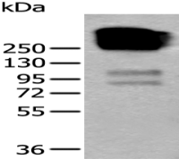
Collagen VII
- 抗体名:
7型胶原抗体
- 适应物种:
人/动物/植物
- 应用范围:
WB,ELISA等
- 浓度:
1mg/ml
- 保存条件:
-20 °
- 规格:
50ul/100ul/200ul
| 规格: | 50ul | 产品价格: | ¥880.0 |
|---|---|---|---|
| 规格: | 100ul | 产品价格: | ¥1580.0 |
| 规格: | 200ul | 产品价格: | ¥2480.0 |
英文名称 : Collagen VII
中文名称 : 7型胶原抗体
别 名 : Collagen Type VII; Alpha 1 type VII collagen; COL7A1; Collagen alpha 1(VII) chain; Collagen type VII alpha 1; Collagen VII alpha 1 polypeptide; CollagenVII; EBD 1; EBD1; EBDCT; EBR 1; EBR1; LC collagen; Long chain collagen.
研究领域 : 免疫学
抗体来源 : Rabbit
克隆类型 : Polyclonal
交叉反应 : Human, Mouse, Rat, Dog, Pig, Cow, Rabbit,
产品应用 : ELISA=1:500-1000 IHC-P=1:400-800 IHC-F=1:400-800 IF=1:100-500 (石蜡切片需做抗原修复)
not yet tested in other applications.
optimal dilutions/concentrations should be determined by the end user.
分 子 量 : 322kDa
细胞定位 : 细胞外基质
性 状 : Lyophilized or Liquid
浓 度 : 1mg/ml
免 疫 原 : KLH conjugated synthetic peptide derived from human Collagen VII:2801-2944/2944
亚 型 : IgG
纯化方法 : affinity purified by Protein A
储 存 液 : 0.01M TBS(pH7.4) with 1% BSA, 0.03% Proclin300 and 50% Glycerol.
保存条件 : Store at -20 °C for one year. Avoid repeated freeze/thaw cycles. The lyophilized antibody is stable at room temperature for at least one month and for greater than a year when kept at -20°C. When reconstituted in sterile pH 7.4 0.01M PBS or diluent of antibody the antibody is stable for at least two weeks at 2-4 °C.
PubMed : PubMed
产品介绍 : This gene encodes the alpha chain of type VII collagen. The type VII collagen fibril, composed of three identical alpha collagen chains, is restricted to the basement zone beneath stratified squamous epithelia. It functions as an anchoring fibril between the external epithelia and the underlying stroma. Mutations in this gene are associated with all forms of dystrophic epidermolysis bullosa. In the absence of mutations, however, an acquired form of this disease can result from an autoimmune response made to type VII collagen.
Function:
Stratified squamous epithelial basement membrane protein that forms anchoring fibrils which may contribute to epithelial basement membrane organization and adherence by interacting with extracellular matrix (ECM) proteins such as type IV collagen.
Subunit:
Homotrimer. Interacts with MIA3/TANGO1; facilitating its loading into transport carriers and subsequent secretion.
Subcellular Location:
Secreted, extracellular space, extracellular matrix, basement membrane.
Post-translational modifications:
Prolines at the third position of the tripeptide repeating unit (G-X-Y) are hydroxylated in some or all of the chains.
DISEASE:
Note=Epidermolysis bullosa acquisita (EBA) is an autoimmune acquired blistering skin disease resulting from autoantibodies to type VII collagen.
Epidermolysis bullosa dystrophica, autosomal dominant (DDEB) [MIM:131750]: A group of autosomal dominant blistering skin diseases characterized by tissue separation which occurs below the dermal-epidermal basement membrane at the level of the anchoring fibrils. Various clinical types with different severity are recognized, ranging from severe mutilating forms to mild forms with limited and localized scarring, and less frequent extracutaneous manifestations. Note=The disease is caused by mutations affecting the gene represented in this entry.
Epidermolysis bullosa dystrophica, autosomal recessive (RDEB) [MIM:226600]: A group of autosomal recessive blistering skin diseases characterized by tissue separation which occurs below the dermal-epidermal basement membrane at the level of the anchoring fibrils. Various clinical types with different severity are recognized, ranging from severe mutilating forms to mild forms with limited and localized scarring, and less frequent extracutaneous manifestations. Mild forms include epidermolysis bullosa mitis and epidermolysis bullosa localisata. Note=The disease is caused by mutations affecting the gene represented in this entry.
Epidermolysis bullosa dystrophica, Pasini type (P-DEB) [MIM:131750]: A severe, dominantly inherited form of dystrophic epidermolysis bullosa characterized by albopapuloid Pasini papule, dorsal extremity blistering, milia formation and red atrophic scarring after recurrent blisters. Note=The disease is caused by mutations affecting the gene represented in this entry.
Epidermolysis bullosa dystrophica, Hallopeau-Siemens type (HS-DEB) [MIM:226600]: The most severe recessive form of dystrophic epidermolysis bullosa. It manifests with mutilating scarring, joint contractures, corneal erosions, esophagus structures, and propensity to formation of cutaneous squamous cell carcinomas leading to premature demise of the affected individuals. Note=The disease is caused by mutations affecting the gene represented in this entry.
Transient bullous dermolysis of the newborn (TBDN) [MIM:131705]: TBDN is a neonatal form of dystrophic epidermolysis bullosa characterized by sub-epidermal blisters, reduced or abnormal anchoring fibrils at the dermo-epidermal junction, and electron-dense inclusions in keratinocytes. TBDN heals spontaneously or strongly improves within the first months and years of life. Note=The disease is caused by mutations affecting the gene represented in this entry.
Epidermolysis bullosa dystrophica, pretibial type (PR-DEB) [MIM:131850]: A form of dystrophic epidermolysis bullosa characterized by pretibial blisters that develop into prurigo-like hyperkeratotic lesions. It predominantly affects the pretibial areas, sparing the knees and other parts of the skin. Other clinical features include nail dystrophy, albopapuloid skin lesions, and hypertrophic scars without pretibial predominance. The phenotype shows considerable interindividual variability. Inheritance is autosomal dominant. Note=The disease is caused by mutations affecting the gene represented in this entry.
Epidermolysis bullosa dystrophica, Bart type (B-DEB) [MIM:132000]: An autosomal dominant form of dystrophic epidermolysis bullosa characterized by congenital localized absence of skin, skin fragility and deformity of nails. Note=The disease is caused by mutations affecting the gene represented in this entry.
Epidermolysis bullosa pruriginosa (EBP) [MIM:604129]: A distinct clinical subtype of epidermolysis bullosa dystrophica. It is characterized by skin fragility, blistering, scar formation, intense pruritus and excoriated prurigo nodules. Onset is in early childhood, but in some cases is delayed until the second or third decade of life. Inheritance can be autosomal dominant or recessive. Note=The disease is caused by mutations affecting the gene represented in this entry.
Nail disorder, non-syndromic congenital, 8 (NDNC8) [MIM:607523]: A nail disorder characterized by isolated toenail dystrophy. The nail changes are most severe in the great toes and consist of the nail plate being buried in the nail bed with a deformed and narrow free edge. Note=The disease is caused by mutations affecting the gene represented in this entry.
Epidermolysis bullosa dystrophica, with subcorneal cleavage (EBDSC) [MIM:131750]: A bullous skin disorder with variable sized clefts just beneath the level of the stratum corneum. Clinical features include blisters, milia, atrophic scarring, nail dystrophy, and oral and conjunctival involvement, as seen in dystrophic epidermolysis bullosa. Note=The disease is caused by mutations affecting the gene represented in this entry.
Similarity:
Contains 1 BPTI/Kunitz inhibitor domain.
Contains 9 fibronectin type-III domains.
Contains 2 VWFA domains.
SWISS:
Q02388
Gene ID:
1294
Important Note:
This product as supplied is intended for research use only, not for use in human, therapeutic or diagnostic applications.
Ⅶ型胶原是胶原蛋白家族中遗传上独特的一个成员,它是锚纤维的主要成分,锚纤维是皮肤基底膜粘着于其下方真皮的复合物.
风险提示:丁香通仅作为第三方平台,为商家信息发布提供平台空间。用户咨询产品时请注意保护个人信息及财产安全,合理判断,谨慎选购商品,商家和用户对交易行为负责。对于医疗器械类产品,请先查证核实企业经营资质和医疗器械产品注册证情况。
 文献和实验
文献和实验叶脉、叶片等分枝形式之一。主要应用于蕨类叶小羽片的生出方式。从主轴分枝的叶脉再分枝时,原来远离主轴基部的一侧,称为上先出型。如复叶耳蕨属Arachnoides(Rumohum)与此相反,称为下先出型,如鳞毛蕨属(Dryopteris)、沼泽蕨属(The-lypteris)。
在对种群的个体分布方式和群落的种数- 个体数关系等进行统计处理时,其随机总体( stochasticpopulation )的类型,称为分布型。总体的类型决定于变量( variate )及其分布函数( distributionfunction )。在变量上有覆盖度和重量有连续( con- tinuous )分布型及个体数有离散( discrete )分布型。连续量分布的基本模型属正态分布( normaldistribution )。当与其不相适应时,多把变量改变为平方根或对数,而以正态
小鼠模型提前制作完成啦!距 CNS 又近了一步!心情也好了一些! 可交付的小鼠基因型对不对怎么确认呢? 之前听说过 PCR 鉴定!还听说过 Southern blot 鉴定!还有没有其他鉴定方式? 不同的鉴定方法都有哪些局限性?基因型鉴定都有哪些坑等着自己跳? 一份小鼠基因型鉴定严谨的标准是什么样子的? 看完今天的介绍你就全部明白了! 常用基因型鉴定包括以下几种方法: 一、双臂 PCR 鉴定 双臂 PCR 是基因编辑小鼠模型基因型检测最常用的方法。方法是选取臂外引物与内部引物,以鼠尾


