大家都在搜

手机验证

询价列表

暂时没有已询价产品
小鼠心肌细胞CD57BL/6小
鼠细胞
企业认证
000
000132
MCM
否
原代细胞
否
Mouse Neurons-dorsal spinal cord
肝脏系统
否
小鼠
否
咨询销售
mouse
干冰
5-10年
贴壁生长
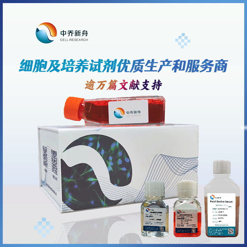
中乔新舟
大量
不是
5 x 10^5 cells/vial
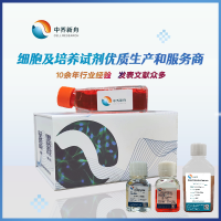
Sciencell公司产品由上海中乔新舟生物科技有限公司优质供应
美国ScienCell研究实验室(www.sciencellonline.com)成立于1999年,公司总部位于美国加州的圣地亚哥。主要致力
于实验室科研用原代细胞、原代细胞专用培养基、原代细胞无血清培养基、干细胞、干细胞培养基、干细胞无血清培养
基的研究和开发,在全球拥有众多客户。在国内销售15年来,很多老师应用其产品发表了高质量的SCI文章,且有着极高
的文献引用率。凭借着严格的质控和优秀的产品品质,深受广大科研工作者的信赖。
ScienCell研究实验室生产的原代细胞、原代细胞专用培养基都经过了严格的质量控制,细胞纯度可达98%。其中包括21
种人体正常细胞系统,90多种不同细胞类型。大多数细胞在全球唯有ScienCell实验室能够成功分离,产品质量过硬。
确保了实验结果的重复性和连贯性。
请登录Sciencell公司官方网站(www.zqxzbio.com 或www.sciencellonline.com )以确保购买正规公司产品

热烈祝贺上海中乔新舟生物科技有限公司成为美国Sciencell公司在中国正规一级代理商:


Sciencell公司产品由上海中乔新舟生物科技有限公司优质供应
近几年的新药研发成功率在逐年下降,其根本原因之一就是传统的药物筛选系统是建立在只具有30-40%
人类基因群的一系列细胞株上。这样一个有“缺陷型”药物筛选系统所产生的药物用于人体上就会出现许
多致命的弱点和不完整性。新一代药物筛选系统是含有一系列近乎完整的人类基因群的原代细胞株。而利
用这些原代细胞株所甄别和筛选出来的候选药物,其诊治人类疾病的成功几率将大大增加。这样不但大大
节省了新药的开发成本,而且将极大地提高人类的健康质量。这些产品从根本上提高了全球生命医学研究、
人类重要疾病药物研发、新药研发的成功率。
所以上海中乔新舟生物科技有限公司致力于提供优质原代细胞产品和完善的售后服务。
Sciencell公司部分原代细胞目录(如需要其他细胞资料请发邮件至 wwwfudan@163.com索取):
上海中乔新舟专长于为生物医药领域的医疗机构、研究中心、企业、临床医生等提供课题设计、基金联合申请、实验
技术服务、论文相关服务、采购外包等整体服务,目前已经成长为国内转化医学外包品牌。
中乔新舟的PI团队已经发展到专职PI 20人,联席PI 312人,其中大多数拥有海外背景。截止到2015年以PI或联席PI为
第一作者发表SCI 论文1682 篇(总IF 值为5721.82,其中影响因子IF>5.0 的论文216 篇,IF>10.0论文32 篇)。
电话:021-56468627
Email:wwwfudan@163.com zqxzbio@vip.163.com
企业QQ:4000389959 QQ:739782475 1957645861
网址:www.zqxzbio.com
风险提示:丁香通仅作为第三方平台,为商家信息发布提供平台空间。用户咨询产品时请注意保护个人信息及财产安全,合理判断,谨慎选购商品,商家和用户对交易行为负责。对于医疗器械类产品,请先查证核实企业经营资质和医疗器械产品注册证情况。
 技术资料
技术资料
需要更多技术资料 索取更多技术资料
M6200-57.pdf 附 (下载 0 次)