


- ¥1700 - 4000
- GeneTex

- 美国
- GTX101277
- 2025年07月16日
- WB, ICC/IF, IHC-P, IHC-Wm, IHC, IHC (Free Floating)
- Rabbit
- Human, Mouse, Rat, Zebrafish, Pig

企业认证
相关产品推荐更多 >

![USP13 antibody [C2C3], C-term](https://img1.dxycdn.com/2023/0516/050/2572066211091928561-14.jpg!wh200)

万千商家帮你免费找货
0 人在求购买到急需产品
- 详细信息
- 文献和实验
- 技术资料
- 免疫原:
Recombinant protein encompassing a sequence within the center region of human HMGB1. The exact sequence is proprietary.
- 亚型:
IgG
- 形态:
Liquid
- 保存条件:
Store as concentrated solution. Centrifuge briefly prior to opening vial. For short-term storage (1-2 weeks), store at 4ºC. For long-term storage, aliquot and store at -20ºC or below. Avoid multiple freeze-thaw cycles.
- 克隆性:
Polyclonal
- 标记物:
Unconjugated
- 适应物种:
Human, Mouse, Rat, Zebrafish, Pig
- 保质期:
12 months from the shipping date of the product.
- 抗原来源:
Human
- 目录编号:
GTX101277
- 级别:
Primary Antibodies
- 库存:
Available
- 供应商:
GeneTex
- 宿主:
Rabbit
- 应用范围:
WB, ICC/IF, IHC-P, IHC-Wm, IHC, IHC (Free Floating)
- 浓度:
0.46 mg/ml (Please refer to the vial label for the specific concentration.)
- 靶点:
HMGB1
- 抗体英文名:
HMGB1 antibody
- 抗体名:
HMGB1 抗体
- 规格:
100 μl/25 μl
| 规格: | 100 μl | 产品价格: | ¥4000.0 |
|---|---|---|---|
| 规格: | 25 μl | 产品价格: | ¥1700.0 |
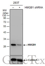
Non-transfected (–) and transfected (+) 293T whole cell extracts (30 μg) were separated by 12% SDS-PAGE, and the membrane was blotted with HMGB1 antibody (GTX101277) diluted at 1:5000. The HRP-conjugated anti-rabbit IgG antibody (GTX213110-01) was used to detect the primary antibody.
HMGB1 antibody detects HMGB1 protein by western blot analysis.
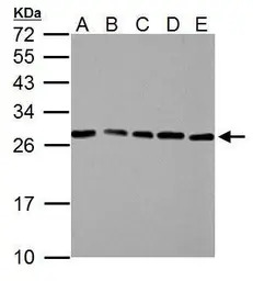
A. 30 μg NIH-3T3 whole cell lysate/extract
B. 30 μg JC whole cell lysate/extract
C. 30 μg BCL-1 whole cell lysate/extract
D. 30 μg C2C12 whole cell lysate/extract
E. 30 μg Raw264.7 whole cell lysate/extract
12% SDS-PAGE
HMGB1 antibody (GTX101277) dilution: 1:3000
The HRP-conjugated anti-rabbit IgG antibody (GTX213110-01) was used to detect the primary antibody.
HMGB1 antibody detects HMGB1 protein at nucleus on mouse colon by immunohistochemical analysis.
Sample: Paraffin-embedded mouse colon.
HMGB1 antibody (GTX101277) dilution: 1:1000.
Antigen Retrieval: Trilogy™ (EDTA based, pH 8.0) buffer, 15min
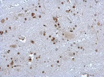
HMGB1 antibody detects HMGB1 protein at nucleus on rat brain stem by immunohistochemical analysis.
Sample: Paraffin-embedded rat brain stem.
HMGB1 antibody (GTX101277) dilution: 1:1000.
Antigen Retrieval: Trilogy™ (EDTA based, pH 8.0) buffer, 15min
HMGB1 antibody detects HMGB1 protein at nucleus in mouse esophagus by immunohistochemical analysis.
Sample: Paraffin-embedded mouse esophagus.
HMGB1 antibody (GTX101277) diluted at 1:1000.
Antigen Retrieval: Citrate buffer, pH 6.0, 15 min
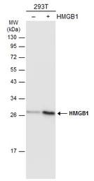
Non-transfected (–) and transfected (+) 293T whole cell extracts (30 μg) were separated by 12% SDS-PAGE, and the membrane was blotted with HMGB1 antibody (GTX101277) diluted at 1:5000. The HRP-conjugated anti-rabbit IgG antibody (GTX213110-01) was used to detect the primary antibody.
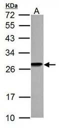
HMGB1 antibody detects HMGB1 protein by western blot analysis.
A. 30 μg PC-12 whole cell lysate/extract
12% SDS-PAGE
HMGB1 antibody (GTX101277) dilution: 1:3000
The HRP-conjugated anti-rabbit IgG antibody (GTX213110-01) was used to detect the primary antibody.
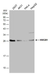
Various whole cell extracts (30 μg) were separated by 12% SDS-PAGE, and the membrane was blotted with HMGB1 antibody (GTX101277) diluted at 1:3000. The HRP-conjugated anti-rabbit IgG antibody (GTX213110-01) was used to detect the primary antibody.
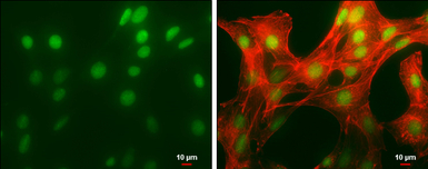
HMGB1 antibody detects HMGB1 protein at nucleus by immunofluorescent analysis.
Sample: NIH/3T3 cells were fixed in 4% PFA at RT for 15 min.
Green: HMGB1 protein stained by HMGB1 antibody (GTX101277) diluted at 1:500.
Red: phalloidin, a cytoskeleton marker, diluted at 1:50.
Scale bar = 10 μm.
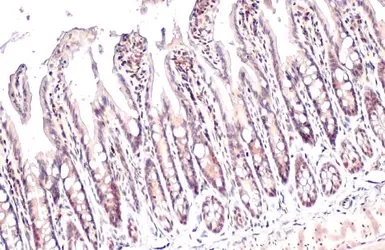
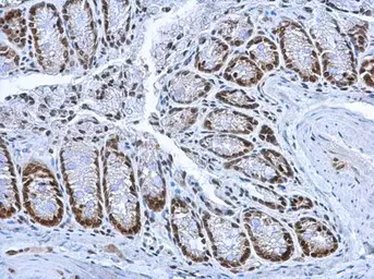
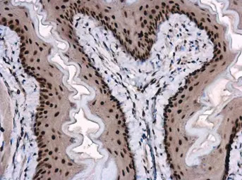
HMGB1 antibody detects HMGB1 protein at nucleus by immunohistochemical analysis.
Sample: Paraffin-embedded mouse intestine.
HMGB1 stained by HMGB1 antibody (GTX101277) diluted at 1:500.
Antigen Retrieval: Citrate buffer, pH 6.0, 15 min
风险提示:丁香通仅作为第三方平台,为商家信息发布提供平台空间。用户咨询产品时请注意保护个人信息及财产安全,合理判断,谨慎选购商品,商家和用户对交易行为负责。对于医疗器械类产品,请先查证核实企业经营资质和医疗器械产品注册证情况。
 文献和实验
文献和实验Huang L et al., Aging (Albany NY) 2019 (PMID:31481647)
Coleman LG Jr et al., Brain Behav Immun 2017 (PMID:29102800)
Masola V et al., FASEB J 2017 (PMID:28970256)
Liu PL et al., PLoS One 2017 (PMID:28727734)
Lin WC et al., RSC Adv. 2017 7()
Lin TJ et al., Mol Cancer 2015 (PMID:26403780)
Lin CY et al., Cell Death Discov 2016 (PMID:27752362)
Dick MS et al., Nat Commun 2016 (PMID:27329339)
Bhatia A et al., Mol Ther Methods Clin Dev 2016 (PMID:27382602)
Sundararaman B et al., Mol Cell 2016 (PMID:26990993)
Juranek JK et al., Front Cell Neurosci 2015 (PMID:26733811)
Ruhl S et al., Eur J Immunol 2015 (PMID:26173909)
Xu YF et al., World J Gastroenterol 2015 (PMID:25805932)
Lee SM et al., Int J Cardiol 2015 (PMID:25939127)
Kayagaki N et al., Nature 2011 (PMID:22002608)
Zhengke Li et al., FASEB J 2021 (PMID:33811702)
Lin C.Y. et al., Natural Product Communications 2021 (Epub)
Sixto-L?pez Y et al., Int J Mol Sci 2020 (PMID:32824279)
D繹rsam B et al., Proc Natl Acad Sci U S A 2018 (PMID:29632181)
Kanno Y et al., Arthritis Res Ther 2020 (PMID:32272967)
Contis-Montes de Oca A et al., Oncotarget 2018 (PMID:30279967)
Fluorescent Biosensors for the Detection of HMGB1 Release
During necrosis and following some instances of apoptosis (in particular in the absence of a proficient phagocytic system), the nonhistone chromatin component high-mobility group box 1 (HMGB1) is released in the extracellular space. In vivo
因为journal club要讲文章 顺便儿拿来和大家讨论下 Hmgb1调节TLR4介导的angiogenesis 最近才开始关注这个蛋白。这篇文章已经在线发表在atvb上,由中山医眼科中心完成的工作。 Hmgb1不仅可以作为一种nuclear factor, 而且还可以作为cytokine来介导感染引起的炎症,免疫损伤。Hmgb1已经在sepsis, arthritis, 肿瘤和其他多种疾病中明确了其作用。Hmgb1可以结合tlr2/4,并且通过MyD88依赖性信号通路促进
Generation of Antibody Molecules Through Antibody Engineering
been overcome to a large extent using genetic-engineering techniques to produce chimeric mouse/human and completely human antibodies. Such an approach is particularly suitable because of the domain structure of the antibody molecule ( 2 ), where functional
 技术资料
技术资料暂无技术资料 索取技术资料

