


- ¥1700 - 4000
- GeneTex

- 美国
- GTX100042
- 2025年07月15日
- WB, ICC/IF, IHC-P, IHC-Fr, FACS, IHC, IHC (Free Floating)
- Rabbit
- Human, Mouse, Rat

企业认证
相关产品推荐更多 >


![TIGIT antibody [1G9] (PE)](https://img1.dxycdn.com/2023/0711/451/6106825198874858761-14.jpg!wh200)
![PTP4A3 antibody [N1C3]](https://img1.dxycdn.com/2023/0711/255/2344116916935858761-14.jpg!wh200)
万千商家帮你免费找货
0 人在求购买到急需产品
- 详细信息
- 文献和实验
- 技术资料
- 免疫原:
Carrier-protein conjugated synthetic peptide encompassing a sequence within the C-terminus region of human Iba1. The exact sequence is proprietary.
- 亚型:
IgG
- 形态:
Liquid
- 保存条件:
Store as concentrated solution. Centrifuge briefly prior to opening vial. For short-term storage (1-2 weeks), store at 4ºC. For long-term storage, aliquot and store at -20ºC or below. Avoid multiple freeze-thaw cycles.
- 克隆性:
Polyclonal
- 标记物:
Unconjugated
- 适应物种:
Human, Mouse, Rat
- 保质期:
12 months from the shipping date of the product.
- 抗原来源:
Human
- 目录编号:
GTX100042
- 级别:
Primary Antibodies
- 库存:
Available
- 供应商:
GeneTex
- 宿主:
Rabbit
- 应用范围:
WB, ICC/IF, IHC-P, IHC-Fr, FACS, IHC, IHC (Free Floating)
- 浓度:
0.06 mg/ml (Please refer to the vial label for the specific concentration.)
- 靶点:
Iba1
- 抗体英文名:
Iba1 antibody
- 抗体名:
Iba1 抗体
- 规格:
100 μl/25 μl
| 规格: | 100 μl | 产品价格: | ¥4000.0 |
|---|---|---|---|
| 规格: | 25 μl | 产品价格: | ¥1700.0 |
Iba1 antibody detects Iba1 protein by immunohistochemical analysis.
Sample: Paraffin-embedded rat tissues.
Iba1 stained by Iba1 antibody (GTX100042) diluted at 1:100.
Antigen Retrieval: Citrate buffer, pH 6.0, 15 min
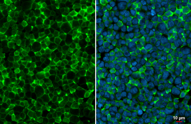
Iba1 antibody detects Iba1 protein at cell membrane by immunofluorescent analysis.
Sample: THP-1 cells were fixed in 4% PFA at RT for 15 min.
Green: Iba1 stained by Iba1 antibody (GTX100042) diluted at 1:500.
Blue: Fluoroshield with DAPI (GTX30920).
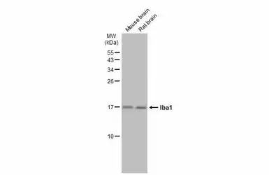
Various tissue extracts (50 μg) were separated by 15% SDS-PAGE, and the membrane was blotted with Iba1 antibody (GTX100042) diluted at 1:1000. The HRP-conjugated anti-rabbit IgG antibody (GTX213110-01) was used to detect the primary antibody.
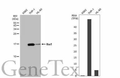
Various whole cell extracts (30 μg) were separated by 15% SDS-PAGE, and the membrane was blotted with Iba1 antibody (GTX100042) diluted at 1:5000. The HRP-conjugated anti-rabbit IgG antibody (GTX213110-01) was used to detect the primary antibody. Correspo
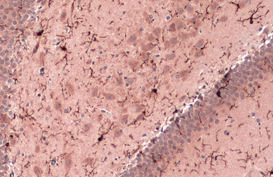
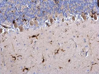
Iba1 antibody detects Iba1 protein at cell membrane and cytoplasm by immunohistochemical analysis.
Sample: Paraffin-embedded rat brain.
Iba1 stained by Iba1 antibody (GTX100042) diluted at 1:500.
Antigen Retrieval: Citrate buffer, pH 6.0, 15 min
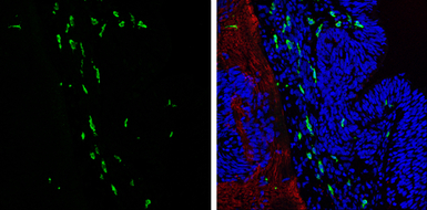
Iba1 antibody detects Iba1 protein expression at microglias by immunohistochemical analysis.
Sample: Frozen sectioned E13.5 Rat brain.
Green: Iba1 protein stained by Iba1 antibody (GTX100042) diluted at 1:250.
Red: beta Tubulin 3/ TUJ1, a mature neuron marker, stained by beta Tubulin 3/ TUJ1 antibody [GT11710] (GTX631836) diluted at 1:500.
Blue: Fluoroshield with DAPI (GTX30920).
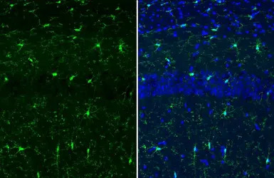
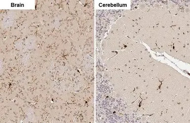
Iba1 antibody detects Iba1 protein at cell membrane and cytoplasm by immunohistochemical analysis.
Sample: Frozen-sectioned mouse brain.
Green: Iba1 stained by Iba1 antibody (GTX100042) diluted at 1:500.
Blue: Fluoroshield with DAPI (GTX30920).
Antigen Retrieval: ice-cold MeOH for 5 min
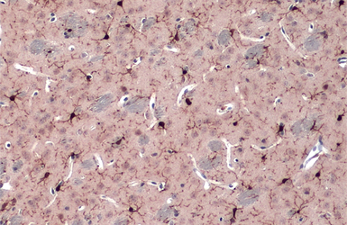
Iba1 antibody detects Iba1 protein at cell membrane and cytoplasm by immunohistochemical analysis.
Sample: Paraffin-embedded mouse brain.
Iba1 stained by Iba1 antibody (GTX100042) diluted at 1:500.
Antigen Retrieval: Citrate buffer, pH 6.0, 15 min
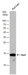
Rat tissue extract (50 μg) was separated by 15% SDS-PAGE, and the membrane was blotted with Iba1 antibody (GTX100042) diluted at 1:1000.
Iba1 antibody detects Iba1 protein on rat hind brain by immunohistochemical analysis.
Sample: Paraffin-embedded rat hind brain.
Iba1 antibody (GTX100042) dilution: 1:500.
Antigen Retrieval: Trilogy™ (EDTA based, pH 8.0) buffer, 15min
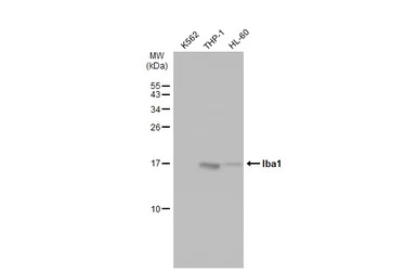
Various whole cell extracts (30 μg) were separated by 15% SDS-PAGE, and the membrane was blotted with Iba1 antibody (GTX100042) diluted at 1:5000. The HRP-conjugated anti-rabbit IgG antibody (GTX213110-01) was used to detect the primary antibody.
风险提示:丁香通仅作为第三方平台,为商家信息发布提供平台空间。用户咨询产品时请注意保护个人信息及财产安全,合理判断,谨慎选购商品,商家和用户对交易行为负责。对于医疗器械类产品,请先查证核实企业经营资质和医疗器械产品注册证情况。
 文献和实验
文献和实验Chen YT et al., Front Immunol 2022 (PMID:36618412)
Colombo G et al., Nat Neurosci 2022 (PMID:36180790)
Chen TF et al., Part Fibre Toxicol 2022 (PMID:35768852)
Tanikawa S et al., Sci Rep 2023 (PMID:36788295)
Xia K et al., Cell Rep Med 2022 (PMID:36270285)
Hong CT et al., Cells 2022 (PMID:36359864)
Md Sadikul Islam et al., BMC Complement Med Ther 2022 (PMID:35752797)
Shaowei Wang et al., Mol Neurodegener 2022 (PMID:35705959)
Yingli Jing et al., Microbiol Spectr 2022 (PMID:35467388)
Yu-En Lin et al., Elife 2021 (PMID:34779396)
Ryoji Amamoto et al., Elife 2022 (PMID:35315776)
Adriana Sofranko et al., Part Fibre Toxicol 2022 (PMID:35337343)
Gemma Manich et al., Front Cell Neurosci 2020 (PMID:33328887)
Mireia Recasens et al., J Neuroinflammation 2021 (PMID:33482848)
Shaowei Wang et al., Mol Neurodegener 2021 (PMID:33863362)
Alessandro Venturino et al., Cell Rep 2021 (PMID:34233180)
Sheng-Han Lee et al., PLoS One 2021 (PMID:34437570)
Margaret E Maes et al., Mol Ther Methods Clin Dev 2021 (PMID:34703843)
Liu XG et al., J Nanobiotechnology 2020 (PMID:33160377)
Huang WY et al., J Neuroinflammation 2022 (PMID:35109859)
Venturino A et al., STAR Protoc 2021 (PMID:34950889)
Su X et al., Ecotoxicol Environ Saf 2021 (PMID:34555716)
Farrher E et al., Neuroimage 2021 (PMID:34592438)
Keerie A et al., Sci Rep 2021 (PMID:34426623)
Couto E Silva A et al., J Neurochem 2021 (PMID:34216036)
Wang M et al., J Neuroinflammation 2021 (PMID:34183019)
Toral-Rios D et al., Int J Mol Sci 2020 (PMID:33050466)
Hara T et al., Cancer Cell 2021 (PMID:34087162)
Chang CW et al., EJNMMI Res 2021 (PMID:33725191)
Kaehler K et al., Invest Ophthalmol Vis Sci 2020 (PMID:32503050)
Ma P et al., J Mol Cell Biol 2021 (PMID:33386850)
Wu CY et al., Prostaglandins Leukot Essent Fatty Acids 2021 (PMID:33445063)
Zuliani I et al., Neurotherapeutics 2020 (PMID:33258073)
Cao K et al., Aging (Albany NY) 2020 (PMID:32003755)
Li H et al., Int J Biol Sci 2020 (PMID:32174788)
Ding B et al., J Neuroinflammation 2020 (PMID:33054814)
Yang L et al., Phytomedicine 2021 (PMID:33091854)
Lee RH et al., Am J Physiol Heart Circ Physiol 2020 (PMID:32946263)
Werner G et al., EMBO Rep 2020 (PMID:32929860)
Di Domenico M et al., Neurotoxicology 2020 (PMID:32450181)
Wahle T et al., Neurochem Int 2020 (PMID:32422323)
Bartsch K et al., Front Immunol 2018 (PMID:29662492)
Patrick L?ningschr?r et al., Cell Rep 2020 (PMID:32160553)
Sozmen M et al., J Comp Pathol 2020 (PMID:32359638)
Zhao Y et al., Neurochem Res 2020 (PMID:32112178)
Petrushina I et al., Neurobiol Dis 2020 (PMID:32119976)
Yu XL et al., Br J Pharmacol 2020 (PMID:32034757)
Patrick Luningschror et al., Cell Rep. 2020;30(10)
Cao K et al., Alzheimers Res Ther 2019 (PMID:31010414)
Hong Zeng et al., Journal of Neuroinflammation 2019 (Epub)
Hong Li et al., bioRxiv 2019 (Epub)
Olechnowicz SWZ et al., Sci Rep 2019 (PMID:31578352)
Pejman S et al., Brain Res Bull 2019 (PMID:31589901)
Bai KJ et al., Chem Biol Interact 2019 (PMID:31348917)
Zhang T et al., FASEB J 2019 (PMID:31365278)
Liao KH et al., Brain Res 2019 (PMID:31276640)
Marques ARA et al., Autophagy 2019 (PMID:31282275)
G?tzl JK et al., EMBO Mol Med 2019 (PMID:31122931)
Kamarehei M et al., Brain Res Bull 2019 (PMID:30738137)
Wahl D et al., Cell Rep 2018 (PMID:30463018)
Huang YR et al., Neurobiol Dis 2018 (PMID:30481547)
Josephine Juettner et al., bioRxiv 2018 (Epub)
Jhan MK et al., J Leukoc Biol 2018 (PMID:30044892)
Ji M et al., Alzheimers Res Ther 2018 (PMID:29914543)
Wolf H et al., Dis Model Mech 2016 (PMID:27491075)
Liu SY et al., Brain Res 2018 (PMID:29902468)
Huang WY et al., J Neuroinflammation 2018 (PMID:29753328)
Hashiguchi T et al., Drug Metab Dispos 2018 (PMID:29626075)
Huang YN et al., Sci Rep 2018 (PMID:29402897)
Souza LC et al., Mol Cell Neurosci 2018 (PMID:29369791)
Lin W et al., Cell Transplant 2017 (PMID:29338384)
Ingold I et al., Cell 2017 (PMID:29290465)
Lin HJ et al., Cell Physiol Biochem 2017 (PMID:29227981)
Huang N et al., Oncotarget 2017 (PMID:29113362)
Zera K et al., PLoS One 2017 (PMID:29045486)
Stroobants S et al., Neurobiol Dis 2017 (PMID:28720484)
Wang SW et al., Alzheimers Res Ther 2017 (PMID:28592267)
Kowalewski B et al., Hum Mol Genet 2015 (PMID:25452429)
Zha J et al., Sci Rep 2016 (PMID:27824125)
Liu HS et al., Sci Rep 2016 (PMID:26750705)
Kowalewski B et al., Hum. Mol. Genet. 2014;24(7)
Lee YZ et al., Int J Mol Sci 2023 (PMID:37047769)
Huang TL et al., Chin Neurosurg J 2022 (PMID:36476392)
Hironao KY et al., Front Nutr 2022 (PMID:36299984)
Bartalska K et al., iScience 2022 (PMID:35789843)
Kiani Shabestari S et al., Cell Rep 2022 (PMID:35705056)
Uchi T et al., J Neuroinflammation 2023 (PMID:36788526)
Cabron AS et al., Acta Neuropathol Commun 2023 (PMID:36707901)
Generation of Antibody Molecules Through Antibody Engineering
been overcome to a large extent using genetic-engineering techniques to produce chimeric mouse/human and completely human antibodies. Such an approach is particularly suitable because of the domain structure of the antibody molecule ( 2 ), where functional
The importance of antibody molecules was first recognized in the 1890s, when it was shown that immunity to tetanus and diphtheria was caused by antibodies against the bacterial exotoxins (1 ). Around the same time, it was shown that antisera
General comments: Antibodies, like most proteins, do not like to be freeze-thawed. Avoid repetitive freezing of your solution. The best way to store your antibody is to keep a high protein concentration (>1 mg/ml), add some protease
 技术资料
技术资料暂无技术资料 索取技术资料

