


- ¥1700 - 4000
- GeneTex

- 美国
- GTX122148
- 2025年07月15日
- WB, ICC/IF, IHC-P, IP, ChIP assay, IHC
- Rabbit
- Human, Mouse, Rat, Drosophila, Pig, Monkey, Fungi, Golden Syrian Hamster, Rice

企业认证
相关产品推荐更多 >

![TNF alpha antibody [TNF/1500R]](https://img1.dxycdn.com/2023/0711/451/6106825198874858761-14.jpg!wh200)
万千商家帮你免费找货
0 人在求购买到急需产品
- 详细信息
- 文献和实验
- 技术资料
- 免疫原:
Carrier-protein conjugated synthetic peptide encompassing a sequence within the N-terminus region of human Histone H3. The exact sequence is proprietary.
- 亚型:
IgG
- 形态:
Liquid
- 保存条件:
Store as concentrated solution. Centrifuge briefly prior to opening vial. For short-term storage (1-2 weeks), store at 4ºC. For long-term storage, aliquot and store at -20ºC or below. Avoid multiple freeze-thaw cycles.
- 克隆性:
Polyclonal
- 标记物:
Unconjugated
- 适应物种:
Human, Mouse, Rat, Drosophila, Pig, Monkey, Fungi, Golden Syrian Hamster, Rice
- 保质期:
12 months from the shipping date of the product.
- 抗原来源:
Human
- 目录编号:
GTX122148
- 级别:
Primary Antibodies
- 库存:
Available
- 供应商:
GeneTex
- 宿主:
Rabbit
- 应用范围:
WB, ICC/IF, IHC-P, IP, ChIP assay, IHC
- 浓度:
0.1 mg/ml (Please refer to the vial label for the specific concentration.)
- 靶点:
Histone H3
- 抗体英文名:
Histone H3 antibody
- 抗体名:
Histone H3 抗体
- 规格:
100 μl/25 μl
| 规格: | 100 μl | 产品价格: | ¥4000.0 |
|---|---|---|---|
| 规格: | 25 μl | 产品价格: | ¥1700.0 |
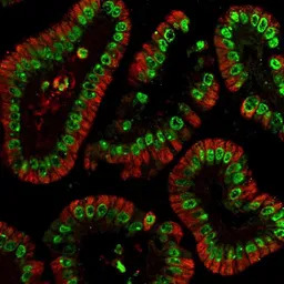
Histone H3 antibody detects Histone H3 protein at nucleus in mouse colon by immunohistochemical analysis.
Sample: Paraffin-embedded mouse colon.
Green: Histone H3 antibody (GTX122148) diluted at 1:500.
Red: alpha Tubulin antibody [GT114] (GTX628802) diluted at 1:500.
Antigen Retrieval: Citrate buffer, pH 6.0, 15 min
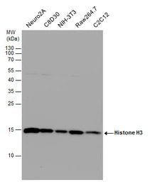
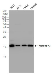
Histone H3 antibody detects Histone H3 protein by western blot analysis. Various whole cell extracts (30 μg) were separated by 15% SDS-PAGE, and the membrane was blotted with Histone H3 antibody (GTX122148) diluted at a dilution of 1:10000. The HRP-conjugated anti-rabbit IgG antibody (GTX213110-01) was used to detect the primary antibody.
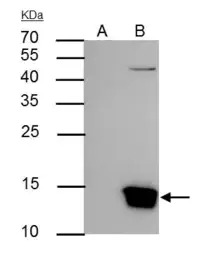
Histone H3 antibody immunoprecipitates Histone H3 protein in IP experiments. IP Sample: Raji whole cell lysate/extract A : Control with 3 μg of pre-immune rabbit IgG B : Immunoprecipitation of Histone H3 by 3 μg of Histone H3 antibody (GTX122148) 15% SDS-PAGE The immunoprecipitated Histone H3 protein was detected by Histone H3 antibody (GTX122148) diluted at 1:5000. EasyBlot anti-rabbit IgG (HRP) (GTX221666-01) was used as a secondary reagent.
Histone H3 antibody detects Histone H3 protein at nucleus in mouse colon by immunohistochemical analysis.
Sample: Paraffin-embedded mouse colon.
Histone H3 antibody (GTX122148) diluted at 1:500.
Antigen Retrieval: Citrate buffer, pH 6.0, 15 min
Histone H3 antibody detects Histone H3 protein by Western blot analysis. Various whole cell extracts (30 μg) were separated by 15% SDS-PAGE, and the membrane was blotted with Histone H3 antibody (GTX122148) diluted at a dilution of 1:1000.
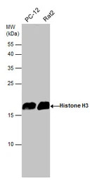
Various whole cell extracts (30 μg) were separated by 15% SDS-PAGE, and the membrane was blotted with Histone H3 antibody (GTX122148) diluted at 1:3000. The HRP-conjugated anti-rabbit IgG antibody (GTX213110-01) was used to detect the primary antibody.
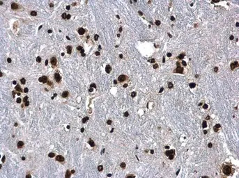
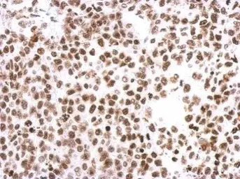
Histone H3 antibody detects Histone H3 protein at nucleus in mouse brain by immunohistochemical analysis.
Sample: Paraffin-embedded mouse brain.
Histone H3 antibody (GTX122148) diluted at 1:500.
Antigen Retrieval: Citrate buffer, pH 6.0, 15 min
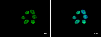
Histone H3 antibody detects Histone H3 protein at nucleus by immunofluorescent analysis.
Sample: A431 cells were fixed in ice-cold MeOH for 5 min.
Green: Histone H3 protein stained by Histone H3 antibody (GTX122148) diluted at 1:500.
Blue: Hoechst 33342 staining.
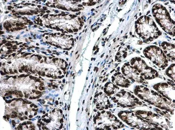
Immunohistochemical analysis of paraffin-embedded Hela xenograft, using Histone H3(GTX122148) antibody at 1:500 dilution.
Antigen Retrieval: Trilogy™ (EDTA based, pH 8.0) buffer, 15min
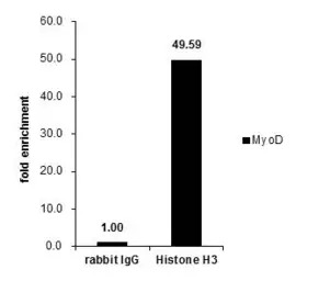
Cross-linked ChIP was performed with HeLa chromatin extract and 5 μg of either control rabbit IgG or anti-Histone H3 antibody. The precipitated DNA was detected by PCR with primer set targeting to MyoD.
风险提示:丁香通仅作为第三方平台,为商家信息发布提供平台空间。用户咨询产品时请注意保护个人信息及财产安全,合理判断,谨慎选购商品,商家和用户对交易行为负责。对于医疗器械类产品,请先查证核实企业经营资质和医疗器械产品注册证情况。
 文献和实验
文献和实验Liu GB et al., Kaohsiung J Med Sci 2020 (PMID:32003536)
Wang D et al., Can J Physiol Pharmacol 2020 (PMID:31935120)
Blin G et al., Nutrients 2020 (PMID:31936461)
Lin YC et al., Int J Mol Sci 2019 (PMID:30934807)
Luc?a D?vila et al., Odontoestomatolog?a 2019 (Epub)
Yi Ding et al., bioRxiv 2019 (Epub)
Dedoni S et al., J Pharmacol Exp Ther 2019 (PMID:31308194)
Lee PC et al., Cancer Cell 2018 (PMID:30537515)
Li M et al., Oncogene 2019 (PMID:30651599)
Hao H et al., Nucleic Acids Res 2018 (PMID:30364964)
Singh R et al., Sci Rep 2018 (PMID:30353146)
Hollingworth R et al., J Virol 2017 (PMID:28855246)
Takashina Y et al., Nutrients 2018 (PMID:30241394)
Lopez-Charcas O et al., Sci Rep 2018 (PMID:30158710)
Fang P et al., Mol Cancer Res 2018 (PMID:29545475)
Chettri P et al., Mol Microbiol 2017 (PMID:29240271)
Lin SC et al., Oncol Rep 2018 (PMID:29207188)
Yang WS et al., PLoS Pathog 2017 (PMID:28212444)
Horke S et al., Infect Immun 2015 (PMID:26056385)
Hii HP et al., PLoS One 2015 (PMID:26218875)
Li PT et al., Int J Mol Sci 2015 (PMID:26473836)
Tenreiro P et al., Anal Biochem 2016 (PMID:27771393)
Shih CC et al., PLoS One 2016 (PMID:27661616)
Li CJ et al., Sci Rep 2015 (PMID:25909282)
Huang YC et al., PLoS Genet 2014 (PMID:25393278)
Lee KH et al., Sci Rep 2014 (PMID:25227736)
Lee YR et al., J Biomed Sci 2012 (PMID:22283874)
Martinez SR et al., Int J Mol Sci 2023 (PMID:37108293)
Y Zhang et al., STAR Protoc 2023 (PMID:37195868)
Tseng KY et al., Cell Death Dis 2023 (PMID:36792604)
Zhao X et al., Front Cell Neurosci 2022 (PMID:36439204)
Zhang Y et al., iScience 2022 (PMID:36185355)
Hao H et al., Nucleic Acids Res 2022 (PMID:35971620)
Yan X et al., Transl Oncol 2022 (PMID:35858494)
Xu Y et al., Cell Biosci 2023 (PMID:36631841)
Lin ZS et al., Elife 2023 (PMID:36622753)
Simona Dedoni et al., Int J Mol Sci 2022 (PMID:35409209)
Simona Dedoni et al., Int J Mol Sci 2021 (PMID:34360553)
Xiuying Sun et al., Bioengineered 2022 (PMID:35333690)
Lei Wang et al., Ann Transl Med 2022 (PMID:35434046)
Alice Migazzi et al., Cell Rep 2021 (PMID:33852844)
Pei-Yi Chu et al., Int J Mol Sci 2021 (PMID:34065390)
Li JN et al., Cancers (Basel) 2021 (PMID:34944924)
Chen J et al., Cell Stress Chaperones 2021 (PMID:33245515)
Li Y et al., Folia Histochem Cytobiol 2022 (PMID:35038162)
Lin S et al., Bioengineered 2021 (PMID:34823423)
Ortiz-Hernandez GL et al., Cells 2021 (PMID:34685704)
Mondrag?n JA et al., Endocrinol Diabetes Metab 2021 (PMID:34505421)
Pang B et al., Cell Biol Int 2021 (PMID:34296787)
Su X et al., Exp Cell Res 2021 (PMID:34181940)
Liu D et al., Cancer Med 2021 (PMID:33838016)
Lin YC et al., Int J Mol Sci 2020 (PMID:32575412)
Zhang Y et al., Drug Des Devel Ther 2021 (PMID:33654384)
Wang W et al., Cell Oncol (Dordr) 2021 (PMID:33469837)
Song L et al., Exp Anim 2020 (PMID:32336744)
Wang B et al., Dev Neurosci 2020 (PMID:33302269)
Chen CY et al., Int J Mol Sci 2020 (PMID:33396303)
Chang TM et al., Cancer Sci 2020 (PMID:33128283)
Hu J et al., Vet Microbiol 2020 (PMID:33045633)
Li X et al., Int J Neurosci 2020 (PMID:32942936)
Han J et al., J Nat Med 2020 (PMID:32761488)
Yuan Q et al., Lab Invest 2020 (PMID:32514126)
Ohnuma K et al., FEBS Lett 2020 (PMID:32294252)
Yuan Q et al., Cancer Cell Int 2020 (PMID:32256208)
Chen CY et al., Front Oncol 2020 (PMID:32158695)
Detection of Histone H3 Phosphorylation in Cultured Cells and Tissue Sections by Immunostaining
. The protocol described here allows the detection of phosphorylated histones in tissue-cultured cells and tissue sections by fluorescent or bright-field immunostaining analysis. Here we used a serine 10 specific P-histone H3 antibody to determine
FACS-Based Detection of Phosphorylated Histone H3 for the Quantitation of Mitotic Cells
scanner (FACS) is described, based on the presence of an intranuclear antigen present only in mitotic cells, detected using a specific, commercially available antibody. Cell staining and FACS analysis can be done in a single day, making this a rapid
Genome-Wide Measurement of Histone H3 Replacement Dynamics in Yeast
chromatin. Understanding the dynamic behavior of chromatin is of great interest for fields ranging from transcriptional regulation through meiosis and gametogenesis. Here, we describe a protocol for measuring histone replacement rates genome wide
 技术资料
技术资料暂无技术资料 索取技术资料

