


- ¥1700 - 4000
- GeneTex

- 美国
- GTX121184
- 2025年07月15日
- WB, ICC/IF, IHC-P, IHC-Fr, IHC-Wm, Dot, EM
- Rabbit
- Human, Mouse, Rat, Zebrafish

企业认证
相关产品推荐更多 >
![M-CSF Receptor antibody [604B5 2E11] (PE)](https://img1.dxycdn.com/2023/0711/451/6106825198874858761-14.jpg!wh200)


![4-1BBL / CD137L antibody [AT113-2]](https://custom.dxycdn.com/trademd/upload/pic/2011/12/14/1323695851.jpg!small.t.1)
万千商家帮你免费找货
0 人在求购买到急需产品
- 详细信息
- 文献和实验
- 技术资料
- 免疫原:
Carrier-protein conjugated synthetic peptide surrounding tri-methyl Lys27 of human Histone H3. The exact sequence is proprietary.
- 亚型:
IgG
- 形态:
Liquid
- 保存条件:
Store as concentrated solution. Centrifuge briefly prior to opening vial. For short-term storage (1-2 weeks), store at 4℃. For long-term storage, aliquot and store at -20℃ or below. Avoid multiple freeze-thaw cycles.
- 克隆性:
Polyclonal
- 标记物:
Unconjugated
- 适应物种:
Human, Mouse, Rat, Zebrafish
- 保质期:
12 months from the shipping date of the product.
- 抗原来源:
Human
- 目录编号:
GTX121184
- 级别:
Primary Antibodies
- 库存:
Available
- 供应商:
GeneTex
- 宿主:
Rabbit
- 应用范围:
WB, ICC/IF, IHC-P, IHC-Fr, IHC-Wm, Dot, EM
- 浓度:
1 mg/ml(Please refer to the vial label for the specific concentration.)
- 靶点:
Histone H3K27me3 (Tri-methyl Lys27)
- 抗体英文名:
Histone H3K27me3 (Tri-methyl Lys27) antibody
- 抗体名:
Histone H3K27me3 (Tri-methyl Lys27) 抗体
- 规格:
100 μl/25 μl
| 规格: | 100 μl | 产品价格: | ¥4000.0 |
|---|---|---|---|
| 规格: | 25 μl | 产品价格: | ¥1700.0 |
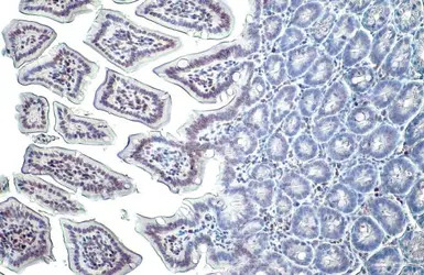
Histone H3K27me3 (trimethyl Lys27) antibody detects Histone H3K27me3 (trimethyl Lys27) protein at nucleus on mouse duodenum by immunohistochemical analysis.
Sample: Paraffin-embedded mouse duodenum.
Histone H3K27me3 (trimethyl Lys27) antibody (GTX121184) diluted at 1:500.
Antigen Retrieval: Trilogy™ (EDTA based, pH 8.0) buffer, 15min

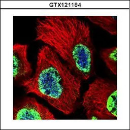
Histone H3K27me3 (Tri-methyl Lys27) antibody detects Histone H3K27me3 (Tri-methyl Lys27) protein at nucleus by immunofluorescent analysis.
Sample: 293T cells were fixed in 4% PFA at RT for 15 min.
Green: Histone H3K27me3 (Tri-methyl Lys27) stained by Histone H3K27me3 (Tri-methyl Lys27) antibody (GTX121184) diluted at 1:500.
Red: alpha Tubulin, a cytoskeleton marker, stained by alpha Tubulin antibody [GT114] (GTX628802) diluted at 1:1000.

Histone H3K27me3 (trimethyl Lys27) antibody detects Histone H3K27me3 (trimethyl Lys27) protein on zebrafish by whole mount immunohistochemical analysis.
Sample: PFA-fixed 2 day-post-fertilization zebrafish embryo.
Histone H3K27me3 (trimethyl Lys27) antibody (GTX121184) dilution: 1:100.
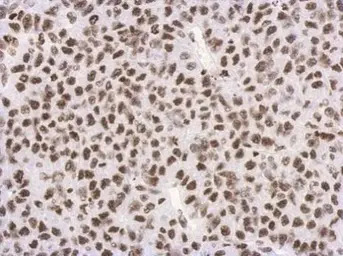
Immunohistochemical analysis of paraffin-embedded Hela xenograft, using Histon H3 (tri-Methyl K27)(GTX121184) antibody at 1:500 dilution.
Antigen Retrieval: Trilogy™ (EDTA based, pH 8.0) buffer, 15min

Histone H3K27me3 (Tri-methyl Lys27) antibody detects Histone H3K27me3 (Tri-methyl Lys27) protein at nucleus by immunofluorescent analysis.
Sample: 293T cells were fixed in 4% PFA at RT for 15 min.
Green: Histone H3K27me3 (Tri-methyl Lys27) stained by Histone H3K27me3 (Tri-methyl Lys27) antibody (GTX121184) diluted at 1:500.
Red: alpha Tubulin, a cytoskeleton marker, stained by alpha Tubulin antibody [GT114] (GTX628802) diluted at 1:1000.
Confocal immunofluorescence analysis (Olympus FV10i) of PFA-fixed A431, using Histone H3 (tri-Methyl K27)(GTX121184) antibody (Green) at 1:500 dilution. Alpha-tubulin filaments were labeled with GTX11304 (Red) at 1:2000.
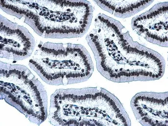
Histone H3K27me3 (Tri-methyl Lys27) antibody detects Histone H3K27me3 (Tri-methyl Lys27) protein at nucleus by immunohistochemical analysis.
Sample: Paraffin-embedded mouse intestine.
Histone H3K27me3 (Tri-methyl Lys27) stained by Histone H3K27me3 (Tri-methyl Lys27) antibody (GTX121184) diluted at 1:500.
Antigen Retrieval: Citrate buffer, pH 6.0, 15 min
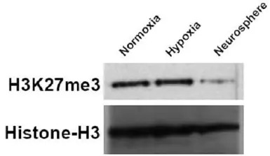
U87 cells were grown under normoxic (DMEM 10% FBS, 16% O2), hypoxic (DMEM 10% FBS, 1% O2), and neurosphere conditions (DMEM/F12, B-27 supplement, growth factor (10ng/ml FGF and 20ng/ml EGF)). Cell lysate were Western blotted for H3K27me3 and Histone-H3 (loading control). The HRP-conjugated anti-rabbit IgG antibody (GTX213110-01) was used to detect the primary antibody.
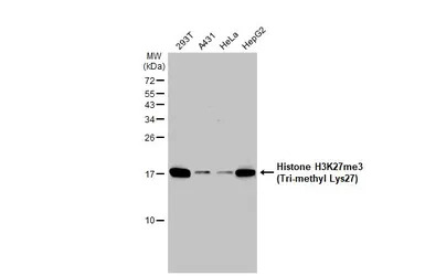
Various whole cell extracts (30 μg) were separated by 15% SDS-PAGE, and the membrane was blotted with Histone H3K27me3 (Tri-methyl Lys27) antibody (GTX121184) diluted at 1:1000. The HRP-conjugated anti-rabbit IgG antibody (GTX213110-01) was used to detect the primary antibody, and the signal was developed with Trident ECL plus-Enhanced.
风险提示:丁香通仅作为第三方平台,为商家信息发布提供平台空间。用户咨询产品时请注意保护个人信息及财产安全,合理判断,谨慎选购商品,商家和用户对交易行为负责。对于医疗器械类产品,请先查证核实企业经营资质和医疗器械产品注册证情况。
 文献和实验
文献和实验Perrigue PM et al., Cancers (Basel) 2020 (PMID:32629974)
Zhang T et al., Int J Nanomedicine 2020 (PMID:32184598)
Hasebe T et al., Gen Comp Endocrinol 2020 (PMID:32084349)
Tanr?kulu B et al., Childs Nerv Syst 2020 (PMID:32025869)
Blin G et al., Nutrients 2020 (PMID:31936461)
Tsuyama N et al., Hematol Oncol 2017 (PMID:28695659)
Mirzamohammadi F et al., Nat Commun 2016 (PMID:27329220)
Perrigue PM et al., Mol Cancer Res 2015 (PMID:25652587)
Liu TP et al., Anticancer Drugs 2015 (PMID:25203626)
Jiang X et al., FASEB J 2012 (PMID:22549509)
upon the dual pulldown to incorporate a third pulldown which is an iteration of the ChIP and is a pulldown for H3K27me3+ (Figure 1b). The third assay described here is the biotin-RNA pulldown of a low-copy RNA that spans the siRNA targeted promoter region
Methyl DNA Immunoprecipitation
can be done. This methylation analysis is highly specific due to the use of a well-characterized monoclonal antibody and each IP is directly quality controlled: two essential keys for reliable results. In addition, the kit protocol is fast and user-friendly. The METHYL kit
Procedure Dilute p27 monoclonal antibody 1:1000 (v/v) in Carbonate Coating Buffer. Add 100 µl/well and incubate o/n @ 4°C or 1 hr @37°C. Wash 3X with TBST (pour onto plate, empty into sink, hit onto towel 3x to clear wells
 技术资料
技术资料暂无技术资料 索取技术资料

