


- ¥1700 - 4000
- GeneTex

- 美国
- GTX113303
- 2025年07月10日
- WB, ICC/IF, IHC-P, IHC-Wm, IP, ChIP assay
- Rabbit
- Human, Mouse, Rat, Zebrafish

企业认证
相关产品推荐更多 >


![Sodium Potassium ATPase antibody [9-A5]](https://img1.dxycdn.com/2023/0711/451/6106825198874858761-14.jpg!wh200)
万千商家帮你免费找货
0 人在求购买到急需产品
- 详细信息
- 文献和实验
- 技术资料
- 免疫原:
Recombinant protein encompassing a sequence within the center region of human HDAC3. The exact sequence is proprietary.
- 亚型:
IgG
- 形态:
Liquid
- 保存条件:
Store as concentrated solution. Centrifuge briefly prior to opening vial. For short-term storage (1-2 weeks), store at 4ºC. For long-term storage, aliquot and store at -20ºC or below. Avoid multiple freeze-thaw cycles.
- 克隆性:
Polyclonal
- 标记物:
Unconjugated
- 适应物种:
Human, Mouse, Rat, Zebrafish
- 保质期:
12 months from the shipping date of the product.
- 抗原来源:
Human
- 目录编号:
GTX113303
- 级别:
Primary Antibodies
- 库存:
Available
- 供应商:
GeneTex
- 宿主:
Rabbit
- 应用范围:
WB, ICC/IF, IHC-P, IHC-Wm, IP, ChIP assay
- 浓度:
0.32 mg/ml (Please refer to the vial label for the specific concentration.)
- 靶点:
HDAC3
- 抗体英文名:
HDAC3 antibody
- 抗体名:
HDAC3 抗体
- 规格:
100 μl/25 μl
| 规格: | 100 μl | 产品价格: | ¥4000.0 |
|---|---|---|---|
| 规格: | 25 μl | 产品价格: | ¥1700.0 |
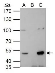
HDAC3 antibody immunoprecipitates HDAC3 protein in IP experiments. IP Sample: 293T whole cell lysate/extract A. 40 μg 293T whole cell lysate/extract B. Control with 2 μg of preimmune rabbit IgG C. Immunoprecipitation of HDAC3 protein by 2 μg of HDAC3 antibody (GTX113303) 7.5% SDS-PAGE The immunoprecipitated HDAC3 protein was detected by HDAC3 antibody (GTX113303) diluted at 1:1000. EasyBlot anti-rabbit IgG (GTX221666-01) was used as a secondary reagent.
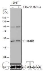
Non-transfected (–) and transfected (+) 293T whole cell extracts (30 μg) were separated by 10% SDS-PAGE, and the membrane was blotted with HDAC3 antibody (GTX113303) diluted at 1:500. The HRP-conjugated anti-rabbit IgG antibody (GTX213110-01) was used to detect the primary antibody.
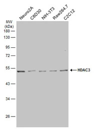
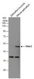
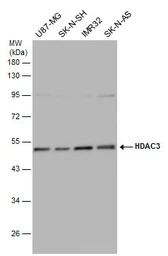
Various whole cell extracts (30 μg) were separated by 10% SDS-PAGE, and the membrane was blotted with HDAC3 antibody (GTX113303) diluted at 1:1000. The HRP-conjugated anti-rabbit IgG antibody (GTX213110-01) was used to detect the primary antibody.
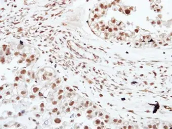
Immunohistochemical analysis of paraffin-embedded human ovarian cancer, using HDAC3(GTX113303) antibody at 1:250 dilution.
Antigen Retrieval: Citrate buffer, pH 6.0, 15 min
Various tissue extracts (30 μg) were separated by 10% SDS-PAGE, and the membrane was blotted with HDAC3 antibody (GTX113303) diluted at 1:500.
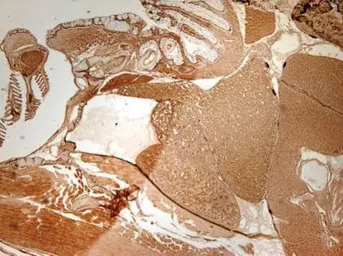
Immunohistochemical analysis of paraffin-embedded zebrafish tissue, using HDAC3 antibody (GTX113303) at 1:300 dilution.
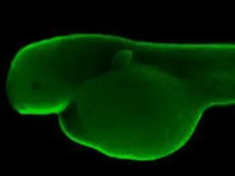
Immunohistochemical analysis (whole mount) of zebrafish embryo, using HDAC3 antibody (GTX113303) at 1:200 dilution.
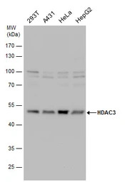
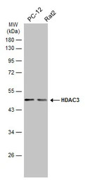
HDAC3 antibody detects HDAC3 protein by Western blot analysis. Various whole cell extracts (30 μg) were separated by 10% SDS-PAGE, and the membrane was blotted with HDAC3 antibody (GTX113303) diluted by 1:1000.
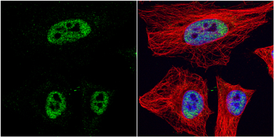
HDAC3 antibody detects HDAC3 protein at nucleus by immunofluorescent analysis.
Sample: HeLa cells were fixed in 4% PFA at RT for 15 min.
Green: HDAC3 protein stained by HDAC3 antibody (GTX113303) diluted at 1:1000.
Red: alpha Tubulin, a cytoskeleton marker, stained by alpha Tubulin antibody [GT114] (GTX628802) diluted at 1:1000.
Blue: Hoechst 33342 staining.
Various whole cell extracts (30 μg) were separated by 10% SDS-PAGE, and the membrane was blotted with HDAC3 antibody (GTX113303) diluted at 1:500. The HRP-conjugated anti-rabbit IgG antibody (GTX213110-01) was used to detect the primary antibody.
Various whole cell extracts (30 μg) were separated by 10% SDS-PAGE, and the membrane was blotted with HDAC3 antibody (GTX113303) diluted at 1:1000. The HRP-conjugated anti-rabbit IgG antibody (GTX213110-01) was used to detect the primary antibody.
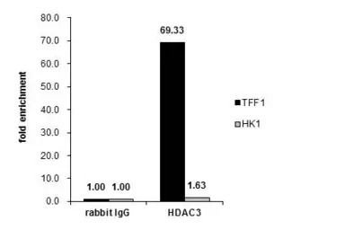
Cross-linked ChIP was performed with MCF-7 chromatin extract and 5 μg of either control rabbit IgG or anti-HDAC3 antibody. The precipitated DNA was detected by PCR with primer set targeting to TFF1 or HK1.
风险提示:丁香通仅作为第三方平台,为商家信息发布提供平台空间。用户咨询产品时请注意保护个人信息及财产安全,合理判断,谨慎选购商品,商家和用户对交易行为负责。对于医疗器械类产品,请先查证核实企业经营资质和医疗器械产品注册证情况。
 文献和实验
文献和实验Miscianinov V. et al., Molecular Therapy 2018 (Epub)
Armour SM et al., Nat Commun 2017 (PMID:28916805)
Emmett MJ et al., Nature 2017 (PMID:28614293)
Soriano FX et al., PLoS One 2011 (PMID:21695276)
Martirosian V et al., Cell Rep 2021 (PMID:34192534)
Miscianinov V et al., Mol Ther 2018 (PMID:29843955)
Generation of Antibody Molecules Through Antibody Engineering
been overcome to a large extent using genetic-engineering techniques to produce chimeric mouse/human and completely human antibodies. Such an approach is particularly suitable because of the domain structure of the antibody molecule ( 2 ), where functional
The importance of antibody molecules was first recognized in the 1890s, when it was shown that immunity to tetanus and diphtheria was caused by antibodies against the bacterial exotoxins (1 ). Around the same time, it was shown that antisera
General comments: Antibodies, like most proteins, do not like to be freeze-thawed. Avoid repetitive freezing of your solution. The best way to store your antibody is to keep a high protein concentration (>1 mg/ml), add some protease
 技术资料
技术资料暂无技术资料 索取技术资料

